
Recallab › Blog › Don't mix these up for NEET PG: complete vs partial hydatidiform mole
Don't mix these up for NEET PG: complete vs partial hydatidiform mole
A high-yield NEET PG pair. Both moles are paternal excess; what separates them is whether a maternal set, and so a fetus, is present. Once that is clear, the villi, the hormone and the risk all follow.
Register to practise NEET PG obstetrics questions →Free account, no card needed.
Complete vs partial hydatidiform mole: side by side
| Complete mole | Partial mole | |
|---|---|---|
| Genetics | Diploid, usually 46,XX, entirely paternal | Triploid, 69, two paternal sets and one maternal |
| Fetal tissue | Absent within the mole | Present |
| Villi | All swollen and avascular | Only some swollen |
| hCG | Very high | Mildly raised |
| Persistent disease | 15 to 20 per cent | 1 to 5 per cent |
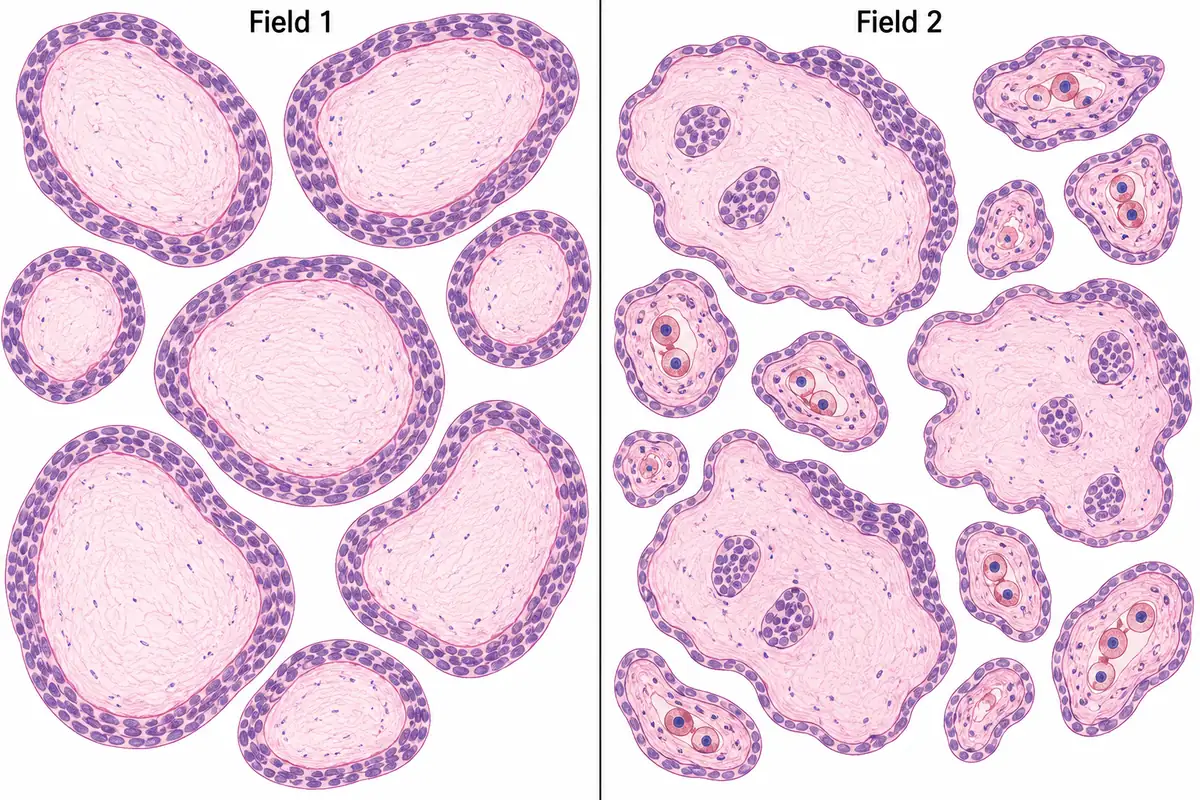
| The clue | No fetal vessels | Nucleated fetal red cells in villous capillaries |
A complete mole forms when an empty ovum is fertilised and the paternal genome duplicates, so there is no maternal material and no embryo. A partial mole forms when two sperm fertilise a normal ovum; the maternal set lets fetal parts and vessels develop. Both need serial hCG follow-up after evacuation.
The one trap
The trap is reaching for central cisterns. They occur in both moles, so they separate neither. The feature that settles it is nucleated fetal red cells in villous capillaries, which only a fetus makes.
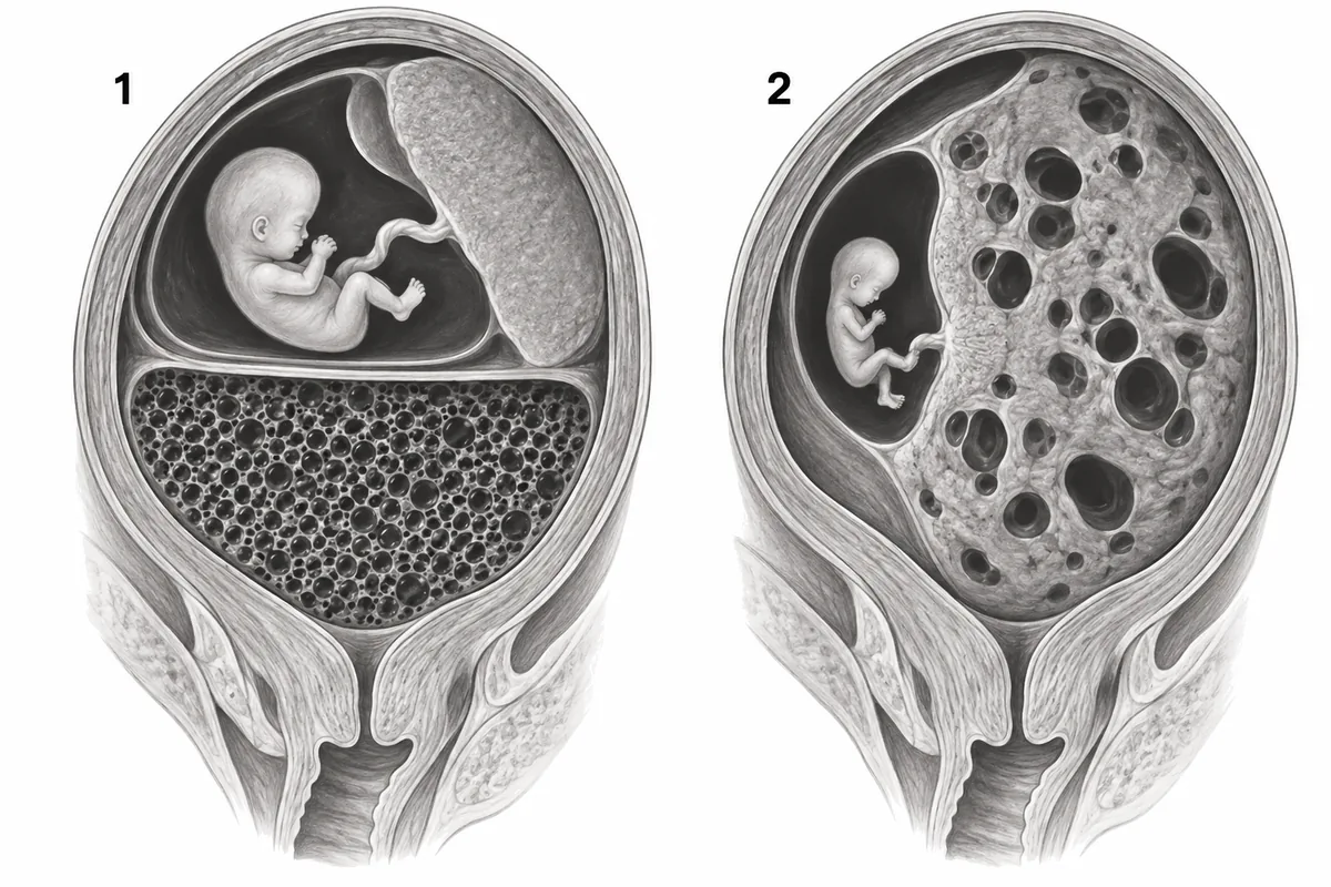
The second trap is a normal fetus beside a complete mole. That may be a coexisting normal twin, with its own placenta, not fetal tissue within the mole.
Try it: say it once before the questions: complete is all father and no fetus; partial has a mother, a fetus and fetal red cells.
The deciding clue
Recallab example 1 · two fields of villi
Two uterine evacuations from women with a bulky uterus and early bleeding are drawn in the figure as histology fields at one magnification. Which single feature in the figure settles which field is a partial mole?
Look first: find the field with two sizes of villus, then look inside the small ones.
 View larger ↗⤢ Tap to enlarge
- AUniform villous swelling
- BScalloped villous outlines
- CCentral cisterns
- DNucleated fetal red cells
Show the answer and explanation
Answer: D. Nucleated fetal red cells
D. Nucleated fetal red cells. The capillaries in field 2’s small villi hold red cells with nuclei, and only a fetus makes those, so field 2 is a partial mole. A complete mole arises from paternal chromosomes alone, so no embryo forms: every villus is swollen, avascular and ringed by trophoblast all round, as in field 1. A partial mole is triploid, with an embryo that lives long enough to make vessels, so only some villi swell and two populations sit side by side.
Why the other options lose: uniform villous swelling (A), with trophoblast all round, is field 1, the complete mole. Scalloped outlines and trapped trophoblast (B) support a partial mole but do not prove a fetus. Central cisterns (C) form in both moles and separate neither.
Recallab question 57571 · Obstetrics › Trophoblastic Disease · reference: Williams Obstetrics
Where the mother shows
Recallab example 2 · a stain that needs the mother
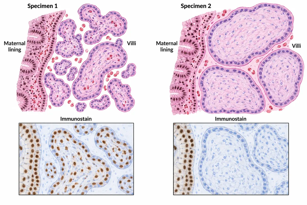
Two uterine evacuation specimens, both showing swollen villi, have each been stained for a protein that a cell can make only from the copy of its gene inherited from the mother. The result is drawn in the figure. What is specimen 2?
Look first: check the maternal lining first: it must stain. Then ask whether the villi do.
 View larger ↗⤢ Tap to enlarge
- AComplete hydatidiform mole
- BChoriocarcinoma
- CPartial hydatidiform mole
- DHydropic abortion
Show the answer and explanation
Answer: A. Complete hydatidiform mole
A. Complete hydatidiform mole. The stained protein, p57, is made only from the maternal copy of its gene. Specimen 2’s villous cells stay unstained because a complete mole has no maternal chromosomes, while the mother’s own lining in the same section stains, proving the stain worked. Specimen 1 stains in both places, so its villi do have a maternal contribution.
Why the other options lose: choriocarcinoma (B) forms no villi, and villi are drawn here. A partial mole (C) carries a maternal set, so its villi stain. A hydropic abortion (D) is a non-molar conception with a maternal set, so its villi stain too.
Recallab question 58520 · Pathology › Female Genital Tract · reference: Robbins, Cotran & Kumar Pathologic Basis of Disease
A fetus beside a mole
Recallab example 3 · two scans at thirteen weeks
Two women at 13 weeks have very high serum hCG and cystic placental tissue on scan. Their scans are drawn in the figure. Which pregnancy may be allowed to continue after counselling?
Look first: count the placentas, then ask where the fetus sits.
 View larger ↗⤢ Tap to enlarge
- ABoth
- BNeither
- CScan 1
- DScan 2
Show the answer and explanation
Answer: C. Scan 1
C. Scan 1. It has two separate placentas, one normal with its fetus and one entirely molar: a complete mole beside a normal twin, with no fetal tissue in the mole. The twin usually has a normal karyotype, which invasive testing confirms, and about four in ten such pregnancies that continue end in a live birth. Continuing does not appear to raise the later risk of trophoblastic neoplasia; pre-eclampsia, bleeding and thyrotoxicosis are watched for, and hCG is followed afterwards.
Why the other options lose: scan 2 is a partial mole, one placenta with focal cysts and a small fetus, whose fetus is usually triploid and rarely survives, so both (A) and scan 2 (D) lose. Neither (B) is wrong because a complete mole beside a normal twin may continue safely.
Recallab question 57877 · Obstetrics › Trophoblastic Disease · reference: Williams Obstetrics
Practise more NEET PG questions →, or watch one-minute teaching shorts in Scroll inside the app.
Why candidates use Recallab
- Built towards 22,000 — a batch at a time
- Illustrations drawn for the question
- Every option explained, not just the right one
- AI study tools that are live today
- Start free, today
Or see the plans, or try a timed mock section.
Register to practise NEET PG obstetrics questionsFree account, no card needed. Practise with every option explained and a reference on every question.Drawn from questions #57571, #58520, #57877, #50578, #50875 in the Recallab bank, each with its own verified answer and current-edition reference. Last reviewed 31 October 2026.