
Recallab › Blog › Don't mix these up for NEET PG: Crohn disease vs ulcerative colitis
Don't mix these up for NEET PG: Crohn disease vs ulcerative colitis
A high-yield NEET PG pair. Both are chronic inflammatory bowel diseases with diarrhoea, and the questions separate them on three things: where the disease is, how deep it goes, and what surgery can do.
Register to practise NEET PG pathology questions →Free account, no card needed.
Crohn disease vs ulcerative colitis: side by side
| Crohn disease | Ulcerative colitis | |
|---|---|---|
| Distribution | Mouth to anus, skip lesions | Colon and rectum, continuous from the rectum upwards |
| Depth | Transmural | Mucosa and submucosa |
| Granulomas | Non-caseating, in about half | Absent |
| Mucosa | Deep linear ulcers, cobblestoning | Red granular mucosa, pseudopolyps |
| Fistulas and strictures | Common | Rare |
| Smoking | Worsens it | Protective |
| Surgery | Can recur in the remaining bowel | Proctocolectomy cures the bowel disease |
Depth explains the complications. Inflammation through the whole wall of the bowel is what lets Crohn disease form fistulas, strictures, abscesses and perianal disease. Ulcerative colitis stays in the lining, so it bleeds instead, and rectal bleeding is characteristic.
In India, the look-alike that matters most for Crohn disease is intestinal tuberculosis. Caseating granulomas and acid-fast bacilli separate them, and the distinction is consequential, because steroids given for tuberculosis are dangerous.
The one trap
The trap is treating any gap as Crohn disease. Skip lesions are the classic Crohn sign, but ulcerative colitis has recognised exceptions. An isolated periappendiceal caecal patch can accompany otherwise typical distal colitis, and backwash ileitis can carry it a short way into the terminal ileum. Neither alone overturns ulcerative colitis.
The reverse trap is crypt abscesses. They occur in both diseases, so they do not prove ulcerative colitis; how deep the inflammation goes is what separates the two.
Try it: say it once before the questions: Crohn roams the gut, through the whole wall; colitis stays in the colon, in the lining.
Pattern and antibodies
Recallab example 1 · two bowel maps, two antibody cards
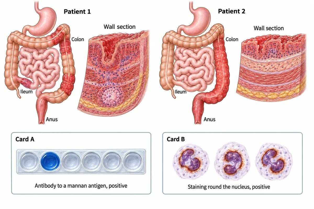
Two patients with months of diarrhoea are drawn in the figure: for each, the bowel actually involved and a section of the wall at the worst point. Beneath them are the two antibody cards from these patients, which have not been matched to them. Which card belongs to patient 1?
Look first: first name each disease from its map and wall section, then match the antibody that leans towards it.
 View larger ↗⤢ Tap to enlarge
- ANeither card
- BCard A
- CBoth cards
- DCard B
Show the answer and explanation
Answer: B. Card A
B. Card A, the antibody to a mannan antigen of yeast (anti-Saccharomyces cerevisiae antibody). Patient 1 has skip lesions, inflammation through the whole wall and a granuloma: Crohn disease, in which this antibody is commoner. Patient 2 has continuous disease from the anus upwards with only the lining involved: ulcerative colitis, in which the perinuclear neutrophil staining pattern is commoner. Both antibodies are missing in many patients, so a negative card proves nothing. They support a diagnosis without settling it; the biopsy decides.
Why the other options lose: neither card (A) ignores that the mannan antibody goes with the patchy transmural disease of patient 1. Both cards (C) would give one patient two antibodies that track different diseases. Card B (D) is the perinuclear pattern, which belongs with the continuous mucosal disease of patient 2.
Recallab question 58316 · Pathology › GIT · reference: Robbins, Cotran & Kumar Pathologic Basis of Disease
The long-standing colon
Recallab example 2 · two contrast enemas
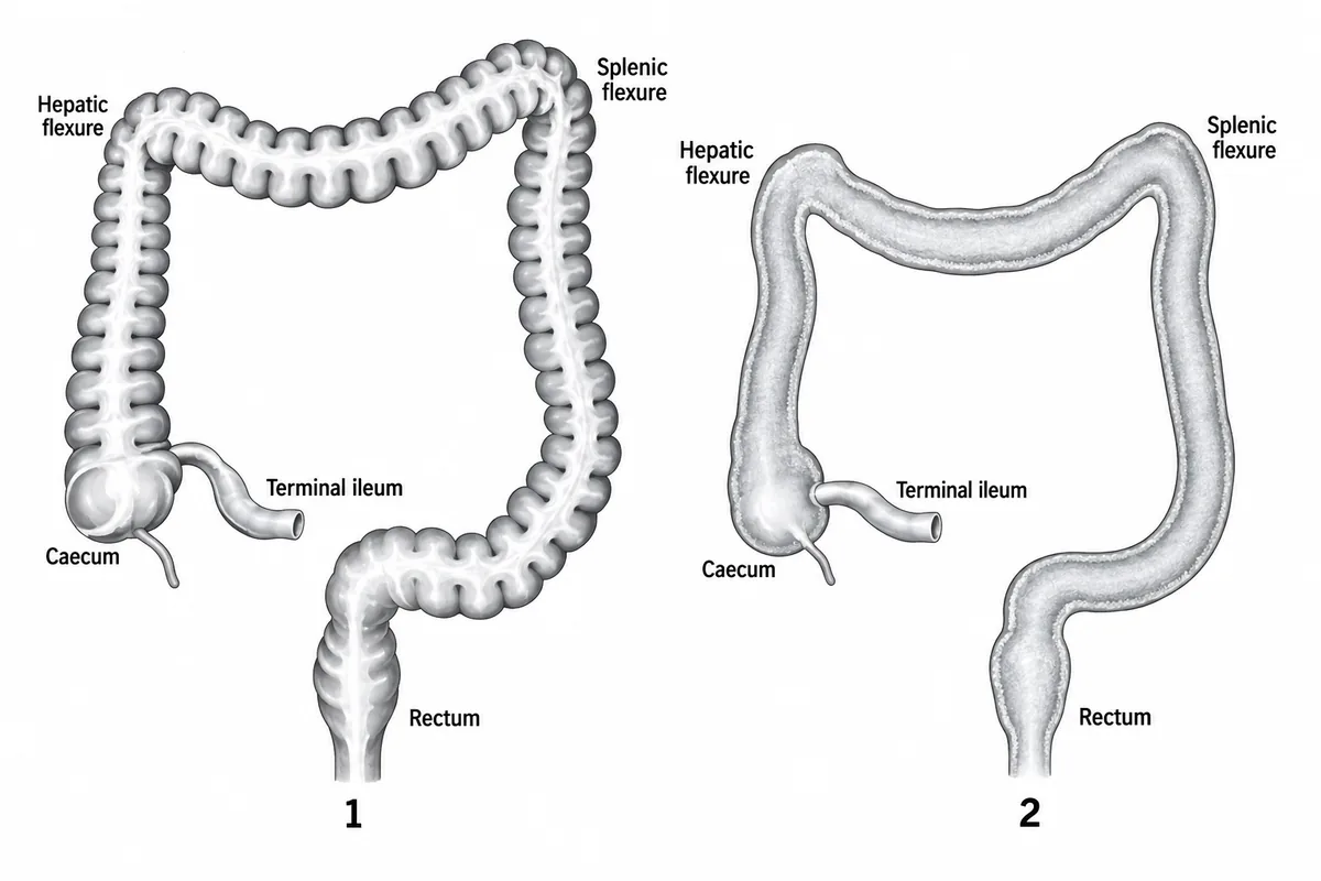
Two contrast enema outlines are drawn in the figure, one from each of two patients who have had diarrhoea for several years. Which feature, rather than the ulceration itself, marks long standing ulcerative colitis?
Look first: compare the walls: which outline has lost its indentations, and does the change run without a break from the rectum?
 View larger ↗⤢ Tap to enlarge
- AShort shouldered narrowing
- BSmooth shortened featureless colon
- CSkip lesions sparing the rectum
- DRegular haustral indentations
Show the answer and explanation
Answer: B. Smooth shortened featureless colon
B. Smooth shortened featureless colon. Outline 2 runs smooth from the rectum to the caecum with no indentations. The haustra are lost because the muscle coat has shortened and thickened, and the flexures sit lower and closer together because the whole colon is shorter. No normal segment interrupts the change, so it is continuous rather than patchy: long-standing ulcerative colitis. Outline 1 is the normal colon, drawn for comparison. The ulceration itself is shown far better at endoscopy.
Why the other options lose: a short shouldered narrowing (A) is the appearance of a tumour. Skip lesions sparing the rectum (C) belong to Crohn disease. Regular haustral indentations (D), as in outline 1, are the normal appearance.
Recallab question 58246 · Radiology › GIT · reference: Bailey & Love's Short Practice of Surgery
What surgery can cure
Recallab example 3 · two colectomy specimens
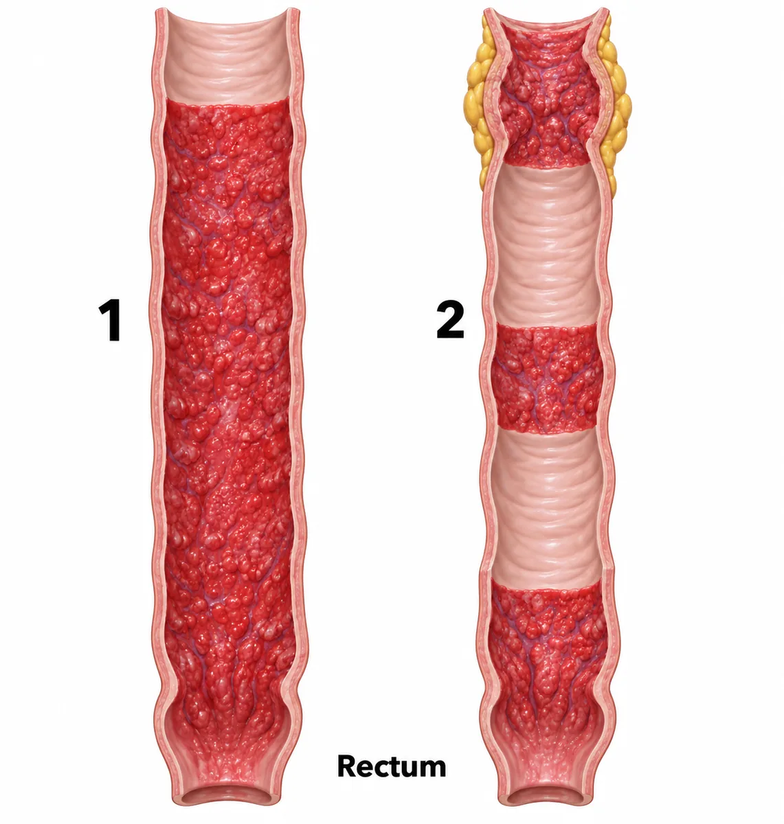
Two resected colons from patients with chronic diarrhoea and pain are drawn in the figure. In which patient does removing the colon and rectum cure the bowel disease?
Look first: name each specimen, then ask which disease stays inside the bowel that was removed.
 View larger ↗⤢ Tap to enlarge
- ANeither patient
- BBoth patients
- CSpecimen 2
- DSpecimen 1
Show the answer and explanation
Answer: D. Specimen 1
D. Specimen 1. Its disease starts at the rectum and runs continuously upwards, with a red granular mucosa and pseudopolyps: ulcerative colitis. Ulcerative colitis is confined to the colon and rectum, apart from backwash ileitis, so removing them removes all the diseased bowel. Specimen 2 has skip lesions, cobblestoning, a thickened wall with a stricture and creeping fat: Crohn disease, which can return anywhere from mouth to anus. The usual operation for ulcerative colitis is a restorative proctocolectomy with an ileal pouch, with a permanent ileostomy when a pouch is not suitable.
Why the other options lose: neither patient (A) is wrong because ulcerative colitis does not return once the colon and rectum are gone. Both patients (B) and specimen 2 (C) are wrong because Crohn disease recurs in the remaining bowel, often at the anastomosis.
Recallab question 55455 · Pathology › GIT & Hepatobiliary Pathology · reference: Robbins, Cotran & Kumar Pathologic Basis of Disease
Practise more NEET PG questions →, or watch one-minute teaching shorts in Scroll inside the app.
Why candidates use Recallab
- Built towards 22,000 — a batch at a time
- Illustrations drawn for the question
- Every option explained, not just the right one
- AI study tools that are live today
- Start free, today
Or see the plans, or try a timed mock section.
Register to practise NEET PG pathology questionsFree account, no card needed. Practise with every option explained and a reference on every question.Drawn from questions #58316, #58246, #55455, #74170, #50123 in the Recallab bank, each with its own verified answer and current-edition reference. Last reviewed 30 October 2026.