
Recallab › Question bank › Dermatology › #50062
Basal cell carcinoma
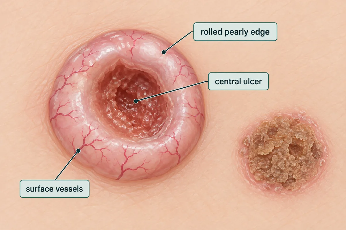
A 68-year-old farmer has a slowly enlarging lesion beside his nose, present for two years. It is a translucent pearly nodule about 8 mm across with a rolled edge, fine telangiectatic vessels crossing the surface, and central ulceration. There is no regional lymphadenopathy. What is the most likely diagnosis?
- correctA. Basal cell carcinoma
- B. Squamous cell carcinoma
- C. Keratoacanthoma
- D. Nodular melanoma
Explanation
Key fact
A pearly rolled edge with surface telangiectasia on sun-exposed facial skin is basal cell carcinoma - locally destructive, virtually never metastatic.
Basal cell carcinoma. The pearly translucent nodule with a rolled edge and telangiectasia is close to diagnostic.
Why
It is on the face, in the sun-exposed area where basal cell carcinoma sits.
The growth has been slow - two years - which fits its indolent behaviour.
A rolled, beaded edge around a central ulcer is the classic rodent ulcer.
The fine vessels running over the surface are characteristic.
No lymphadenopathy, because it spreads locally and almost never metastasises.
A rim you can see round, a centre that sinks
Why not the others
- B. Squamous cell carcinoma — grows faster over months, is keratotic or crusted with an everted edge, and does metastasise to regional nodes.
- C. Keratoacanthoma — erupts over weeks as a dome with a central keratin plug and then often regresses. Two years of slow growth is against it.
- D. Nodular melanoma — is pigmented, grows quickly, and shows asymmetry and colour variation. A translucent pearly nodule is not melanoma.
Remember
Rodent ulcer gnaws, it does not travel. Local destruction can be severe, but nodes stay clear.
Go deeper
The morphoeic or sclerosing variant looks like a scar with indistinct margins and needs a wider excision or Mohs surgery.
Multiple basal cell carcinomas in a young person suggest Gorlin syndrome, from a PTCH1 mutation in the hedgehog pathway.
Fitzpatrick's Dermatology, 9th ed. (McGraw Hill, 2019).