
Recallab › Question bank › ENT › Nose › #50197
Radiographic views of the sinuses
Which plain radiographic view best demonstrates the frontal sinus?
- A. Towne's view
- B. Water's (occipitomental) view
- C. Submentovertical view
- correctD. Caldwell (occipitofrontal) view
Explanation
Key fact
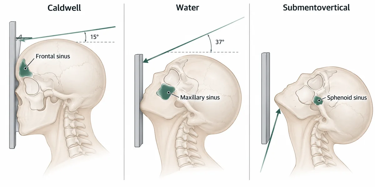
Caldwell for frontal and ethmoid; Water's for maxillary; submentovertical for sphenoid. Each view lifts one sinus clear of the petrous bones.
Caldwell (occipitofrontal) view. The beam is angled to project the petrous ridges below the orbits and display the frontal and ethmoid sinuses.
Why
Plain sinus views differ only in how the head and beam are angled.
The problem in every one is the dense petrous temporal bone.
In the Caldwell view the beam is angled 15 degrees caudally.
That throws the petrous ridges into the lower orbit and clears the frontal sinuses.
The ethmoid air cells are seen between the orbits on the same film.
Which view for which sinus
| View | Position | Best shows |
|---|---|---|
| Caldwell (occipitofrontal) | Nose and forehead to film, beam 15 degrees caudal | Frontal and ethmoid sinuses |
| Water's (occipitomental) | Chin to film, head extended 37 degrees | Maxillary sinuses, orbital floor |
| Submentovertical | Beam under the chin to the vertex | Sphenoid sinus, zygomatic arches |
| Lateral | Side on | Frontal and sphenoid, postnasal space |
Each view clears the petrous bone from one sinus
Why not the others
- A. Towne's view — shows the occipital bone, foramen magnum and mandibular condyles - not the sinuses.
- B. Water's (occipitomental) view — is the maxillary sinus view, and is also the standard view for an orbital blowout fracture. One circulating recall keys it to the frontal sinus, and that is wrong.
- C. Submentovertical view — demonstrates the sphenoid sinus and the zygomatic arches.
Remember
Chin up for maxilla, forehead down for frontal. Water's tilts the chin to the film; Caldwell puts the forehead on it.
Go deeper
Plain films have largely been replaced by computed tomography, which is the standard for sinus disease and is essential before endoscopic sinus surgery.
The maxillary antrum is the first sinus to develop and the last to be fully pneumatised; the frontal sinus is not usually visible on a radiograph until about 6 years of age, which matters when reading a child's film.
Dhingra PL, Dhingra S. Diseases of Ear, Nose and Throat, 9th ed. (Elsevier, 2025).