
Recallab › Question bank › Anatomy › Lower Limbs › #50203
Femoral triangle
A femoral arterial line is being placed. Working from lateral to medial in the femoral triangle just below the inguinal ligament, in what order are the structures encountered?
- A. Artery, nerve, vein, then the femoral canal
- B. Nerve, vein, artery, then the femoral canal
- C. Vein, artery, nerve, then the femoral canal
- correctD. Nerve, artery, vein, then the femoral canal
Explanation
Key fact
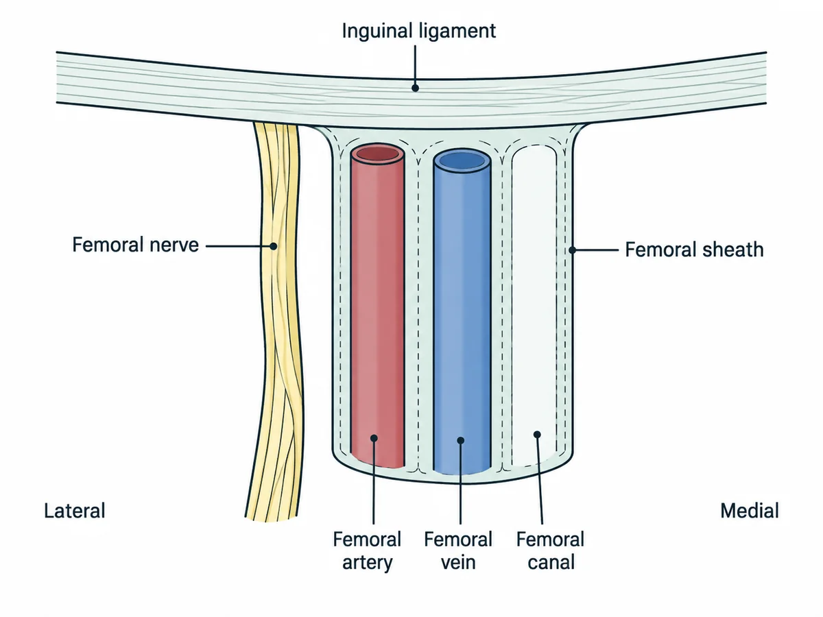
NAVY, lateral to medial: Nerve, Artery, Vein, Y-fronts. The canal, and any femoral hernia, lies medial to everything.
Nerve, artery, vein, then the canal. The pulse is the landmark: the nerve is lateral to it and the vein medial.
Why
The femoral triangle is bounded by the inguinal ligament, sartorius and adductor longus.
The femoral nerve lies outside the femoral sheath, most laterally.
The artery is next, and is the palpable landmark at the mid-inguinal point.
The vein lies immediately medial to it - which is why you aim medial to the pulse for venous access.
The femoral canal, containing fat and a lymph node, is the most medial compartment.
The femoral triangle, lateral to medial
Why not the others
- A. Artery, nerve, vein, then the femoral canal — swaps the first two. The nerve is always lateral to the artery, and it lies outside the sheath.
- B. Nerve, vein, artery, then the femoral canal — swaps artery and vein, so a venous puncture would be attempted where the artery lies.
- C. Vein, artery, nerve, then the femoral canal — reverses the order entirely and would send a venous needle laterally into the nerve.
Remember
NAVY from lateral to medial. Nerve, Artery, Vein, Y-fronts - and the hernia comes out at the Y end.
Go deeper
A femoral hernia passes through the canal, so it appears below and lateral to the pubic tubercle; an inguinal hernia appears above and medial to it.
The femoral nerve is compressed in the puerperium by prolonged lithotomy or a retractor, giving quadriceps weakness and a lost knee jerk.
The common peroneal nerve is the one injured at the neck of the fibula, and causes foot drop - a different nerve at a different site.
Gray's Anatomy for Students, 5th ed. (Elsevier, 2023); Snell's Clinical Anatomy by Regions, 11th ed. (Wolters Kluwer, 2024).