
Recallab › Question bank › Physiology › Respiration › #50210
Alveolar dead space in pulmonary embolism
A patient has a large pulmonary embolus completely obstructing flow to one lung segment, while that segment continues to be ventilated. Compared with the arterial PaCO2, what happens to the end-tidal carbon dioxide?
- correctA. It falls, so the gap between arterial and end-tidal carbon dioxide widens
- B. It falls, and the arterial PaCO2 falls by exactly the same amount
- C. It is unchanged, because ventilation is unchanged
- D. It rises, so the gap narrows
Explanation
Key fact
Dead space dilutes the exhaled carbon dioxide. A widened arterial to end-tidal gradient is the physiological signature of pulmonary embolism.
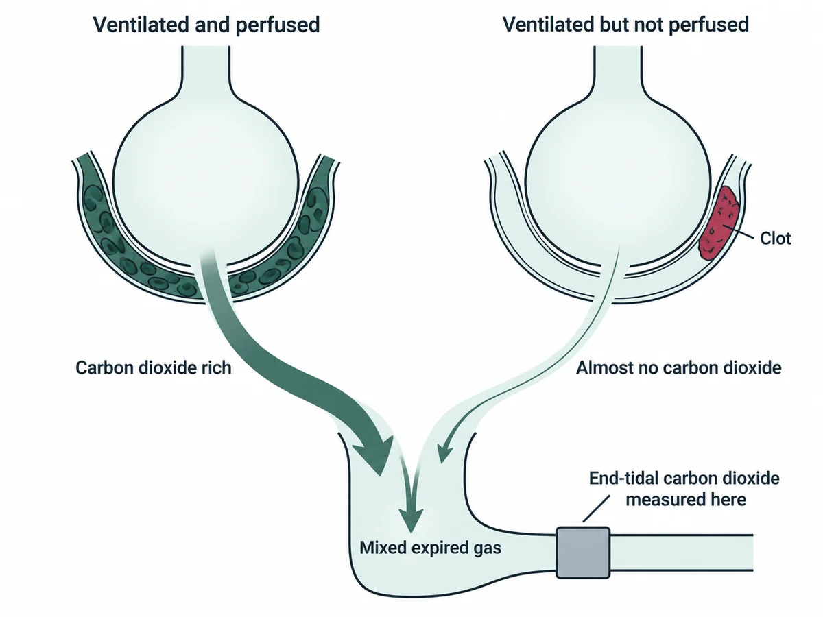
End-tidal carbon dioxide falls and the gradient widens. The obstructed segment contributes gas with almost no carbon dioxide in it.
How it happens
- Carbon dioxide reaches the alveolus only from blood.
- The embolised segment is ventilated but not perfused - alveolar dead space.
- Its gas therefore resembles inspired air, with almost no carbon dioxide.
- That gas mixes with normal alveolar gas during expiration.
- The mixture is diluted, so end-tidal carbon dioxide falls.
- Arterial PaCO2 comes from perfused lung and is normal or even low from hyperventilation, so the gradient widens.
Why dead space lowers end-tidal carbon dioxide
Why not the others
- B. It falls, and the arterial PaCO2 falls by exactly the same amount — would leave the gradient unchanged. In practice arterial carbon dioxide is maintained or lowered by hyperventilation, and the two diverge.
- C. It is unchanged, because ventilation is unchanged — misses the point: the carbon dioxide content of exhaled gas depends on perfusion, not on ventilation alone.
- D. It rises, so the gap narrows — is the opposite. A rise in end-tidal carbon dioxide means more perfusion, not less.
Remember
Shunt lowers oxygen; dead space lowers exhaled carbon dioxide. Two mismatches, two different bedside signatures.
Go deeper
The same reasoning explains why end-tidal carbon dioxide falls abruptly during a cardiac arrest and rises again with return of spontaneous circulation - it is a perfusion monitor as much as a ventilation monitor.
Physiological dead space can be estimated with the Bohr equation, which compares arterial and mixed expired carbon dioxide.
Clinically, the diagnosis still rests on a Wells score with D-dimer or computed tomography pulmonary angiography; the capnography change supports it but is not specific.
Guyton and Hall Textbook of Medical Physiology, 15th ed. (Elsevier, 2026); Harrison's Principles of Internal Medicine, 22nd ed. (McGraw Hill, 2025).