
Recallab › Question bank › Surgery › #50265
Phyllodes tumour
A 42-year-old woman has a 7 cm mobile breast lump that has doubled in size over four months. The overlying skin is stretched and shiny with dilated veins but is not tethered, and there is no axillary lymphadenopathy. Core biopsy shows a leaf-like architecture with clefted epithelium over a hypercellular stroma. What is the diagnosis?
- A. Invasive ductal carcinoma
- B. Fibroadenoma
- C. Breast abscess
- correctD. Phyllodes tumour
Explanation
Key fact
Phyllodes is a fibroadenoma-like lesion that grows fast and needs a wide margin. The stroma, not the epithelium, is the neoplastic part.
Phyllodes tumour. Rapid growth of a large mobile lump with leaf-like architecture and a cellular stroma.
Why
It arises from intralobular stroma, so it is a fibroepithelial tumour.
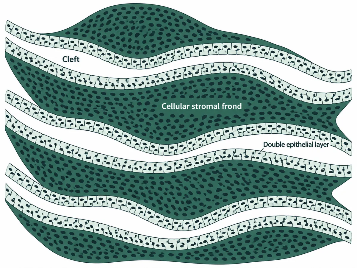
The stroma proliferates and pushes the epithelium into leaf-like clefts - the name means leaf.
Growth is rapid, unlike a fibroadenoma, and the lesion reaches a large size.
Stretched shiny skin with dilated veins reflects pressure, not invasion, so the skin is not tethered.
Spread, when it happens, is haematogenous to the lung - so nodes are usually clear.
The leaf-like architecture of a phyllodes tumour
Why not the others
- A. Invasive ductal carcinoma — characteristically tethers the skin, is hard and irregular rather than mobile, and spreads to axillary nodes.
- B. Fibroadenoma — is usually smaller, occurs in younger women, grows slowly and has a hypocellular stroma without the leaf-like clefting.
- C. Breast abscess — is painful, hot and tender with systemic signs, usually in a lactating woman.
Remember
A fibroadenoma that suddenly grows, in a woman in her forties. That history is phyllodes until the histology says otherwise.
Go deeper
It is graded benign, borderline or malignant on stromal cellularity, atypia, mitoses and the tumour border. Even benign lesions recur if incompletely excised.
Treatment is wide local excision with a margin of at least 1 cm; axillary clearance is not indicated because spread is haematogenous.
Core biopsy can be difficult to interpret, and the distinction from a cellular fibroadenoma sometimes needs the whole excised specimen.
Bailey & Love's Short Practice of Surgery, 28th ed. (CRC Press, 2023); Robbins, Cotran & Kumar Pathologic Basis of Disease, 11th ed. (Elsevier, 2025).