
Recallab › Question bank › Pathology › #50324
Interstitial lung disease in systemic sclerosis
A woman with diffuse cutaneous systemic sclerosis and anti-topoisomerase I antibodies develops progressive exertional breathlessness. High-resolution computed tomography shows bilateral basal ground-glass change with fine reticulation and little honeycombing. Which histological pattern is most likely?
- A. Diffuse alveolar damage
- correctB. Non-specific interstitial pneumonia
- C. Usual interstitial pneumonia
- D. Organising pneumonia
Explanation
Key fact
Systemic sclerosis lung disease is usually non-specific interstitial pneumonia, not the usual interstitial pattern of idiopathic fibrosis. That difference changes the treatment.
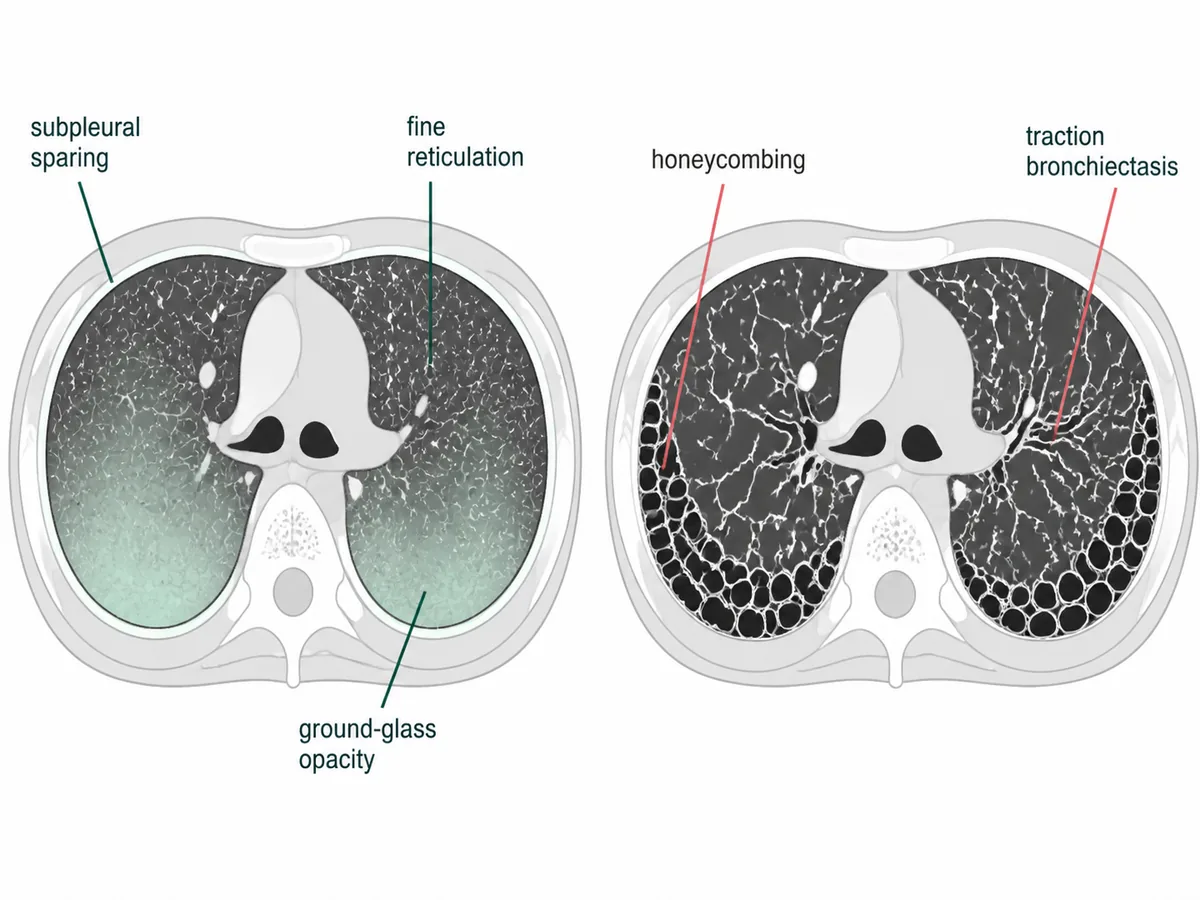
Non-specific interstitial pneumonia. Ground-glass with fine reticulation and little honeycombing is its radiological signature.
Why
The changes are temporally uniform - all areas at the same stage.
That produces ground-glass with fine reticulation rather than coarse honeycombing.
The usual interstitial pattern is patchy in time, with fibroblastic foci.
Anti-topoisomerase I marks the diffuse cutaneous subset, which carries the highest lung risk.
The distinction matters because this pattern often responds to immunosuppression.
Two interstitial patterns compared on high-resolution computed tomography
Why not the others
- A. Diffuse alveolar damage — is the histology of acute respiratory distress syndrome. It develops over days with dense consolidation, not over months.
- C. Usual interstitial pneumonia — is defined by basal subpleural honeycombing with traction bronchiectasis. Honeycombing is conspicuously absent here.
- D. Organising pneumonia — gives patchy peripheral consolidation that migrates between scans, not symmetrical basal reticulation.
Remember
Ground glass without honeycombing points to the treatable pattern.
Go deeper
Mycophenolate and cyclophosphamide both slow decline, and nintedanib is licensed for the progressive fibrosing phenotype in this disease.
Anti-centromere antibodies mark the limited subset, in which pulmonary arterial hypertension rather than fibrosis is the main lung threat.
Screening with pulmonary function tests and computed tomography at diagnosis is recommended, because lung disease is now the leading cause of death.
Harrison's Principles of Internal Medicine, 22nd ed. (McGraw Hill, 2025); Robbins, Cotran & Kumar Pathologic Basis of Disease, 11th ed. (Elsevier, 2025).