
Recallab › Question bank › Orthopaedics › #50373
Posterior dislocation of the hip
A front-seat passenger strikes the dashboard with his flexed knee. The right leg is short, flexed, adducted and internally rotated at the hip. Which nerve is at risk, and which of its divisions is most often affected?
- A. The femoral nerve, with the saphenous branch most affected
- correctB. The sciatic nerve, with the common peroneal division most affected
- C. The obturator nerve, with its anterior division most affected
- D. The sciatic nerve, with the tibial division most affected
Explanation
Key fact
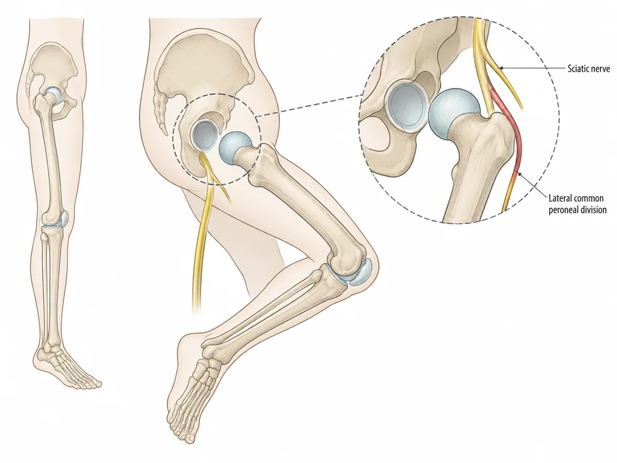
Posterior dislocation puts the femoral head onto the sciatic nerve, and the peroneal division suffers first. Flexed, adducted, internally rotated is the posture.
The sciatic nerve, common peroneal division. Foot drop is the deficit to look for and to document before reduction.
Why
The dashboard drives the femur backwards through a flexed hip and knee.
The head leaves the acetabulum posteriorly, where the sciatic nerve lies.
The peroneal division is lateral, more superficial and has fewer protective fascicles.
It therefore fails first, producing foot drop and loss of dorsiflexion.
The limb rests flexed, adducted and internally rotated.
The dashboard injury and the nerve behind it
Why not the others
- A. The femoral nerve, with the saphenous branch most affected — lies anterior to the hip and is at risk in an anterior dislocation, not a posterior one.
- C. The obturator nerve, with its anterior division most affected — runs through the obturator foramen and is not in the path of a posteriorly displaced head.
- D. The sciatic nerve, with the tibial division most affected — names the right nerve but the wrong division. The tibial part is medial and better protected.
Remember
Posterior: flexed, adducted, internally rotated, and check the foot. Anterior is the mirror image.
Go deeper
This is an orthopaedic emergency: reduction within about six hours markedly reduces the risk of avascular necrosis of the femoral head.
The blood supply comes mainly from the medial circumflex femoral artery through the retinacular vessels, which the dislocation tears.
Computed tomography after reduction is standard, to find acetabular wall fractures and intra-articular fragments that plain films miss.
Apley & Solomon's System of Orthopaedics and Trauma, 10th ed. (CRC Press, 2018); Maheshwari J, Mhaskar VA. Essential Orthopaedics, 8th ed. (Jaypee, 2026).