
Recallab › Question bank › Ophthalmology › #50599
Blow-out fracture
A man is struck in the eye by a cricket ball. He has diplopia on looking up, numbness of the cheek and upper lip, and the globe sits lower than the other side. Which wall has fractured, and what is trapped?
- A. The lateral wall; lateral rectus herniating into the temporal fossa
- B. The orbital roof; superior rectus herniating into the frontal sinus
- C. The medial wall; medial rectus herniating into the sphenoid sinus
- correctD. The orbital floor; inferior rectus and orbital fat herniating into the maxillary sinus
Explanation
Key fact
The floor is the weakest wall and it gives way first. Everything in the presentation follows from what lies beneath it.
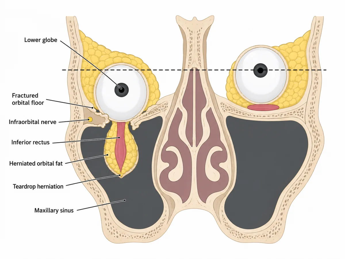
The orbital floor, with inferior rectus and fat herniating into the maxillary sinus. The infraorbital nerve runs in the same floor.
Why
A blunt object larger than the orbital rim raises intraorbital pressure sharply.
The floor is thinnest and fractures, decompressing the orbit downwards.
Inferior rectus and orbital fat prolapse into the maxillary sinus.
Tethering limits upgaze, giving diplopia looking up.
The infraorbital nerve runs in the floor, so the cheek and upper lip go numb.
The blow-out fracture
Why not the others
- A. The lateral wall; lateral rectus herniating into the temporal fossa — is the strongest wall, formed largely by the zygomatic bone and greater wing of sphenoid.
- B. The orbital roof; superior rectus herniating into the frontal sinus — is uncommon and occurs mainly in children; it would not cause infraorbital numbness.
- C. The medial wall; medial rectus herniating into the sphenoid sinus — is thin and does fracture, but into the ethmoid sinus, limiting horizontal gaze and causing surgical emphysema on nose-blowing.
Remember
Floor down, upgaze limited, cheek numb.
Go deeper
The teardrop sign on a coronal computed tomogram is herniated tissue hanging into the maxillary sinus roof.
A white-eyed blow-out in a child with entrapment and an oculocardiac reflex - bradycardia and vomiting on eye movement - is a surgical emergency.
In adults, repair is usually deferred one to two weeks for swelling to settle, and is indicated for persistent diplopia, entrapment or significant enophthalmos.
Khurana AK. Comprehensive Ophthalmology, 10th ed. (Jaypee, 2025); Bailey & Love's Short Practice of Surgery, 28th ed. (CRC Press, 2023).