
Recallab › Blog › Dermatology for NEET PG: six spotters and the clue that settles each
Dermatology for NEET PG: six spotters and the clue that settles each
A skin spotter is solved by looking before naming. Read the edge, the surface and the colour, then ask where the lesion sits and what else the figure shows.
Register to practise NEET PG dermatology questions →Free account, no card needed.
These six illustrated Recallab questions move from a lump on the eyelid to a scaling plaque, a shin, a forehead, a rash that moves and a violet nose. Whether you are learning dermatology in MBBS or revising for NEET PG or INI-CET, try each question before opening its worked answer.
Before you name the lesion: three questions
- What is the edge doing? Look for a rolled rim, a raised coloured border or a trailing edge of scale.
- What are the surface and colour? Pearly, yellow, ivory or violet; flat plates of scale or a cone.
- Where is it, and what else is drawn? The site, a second panel, a second day or a chest outline often decides between look-alikes.
Read the edge, the scale and the colour
Recallab example 1 · four lumps on the lid
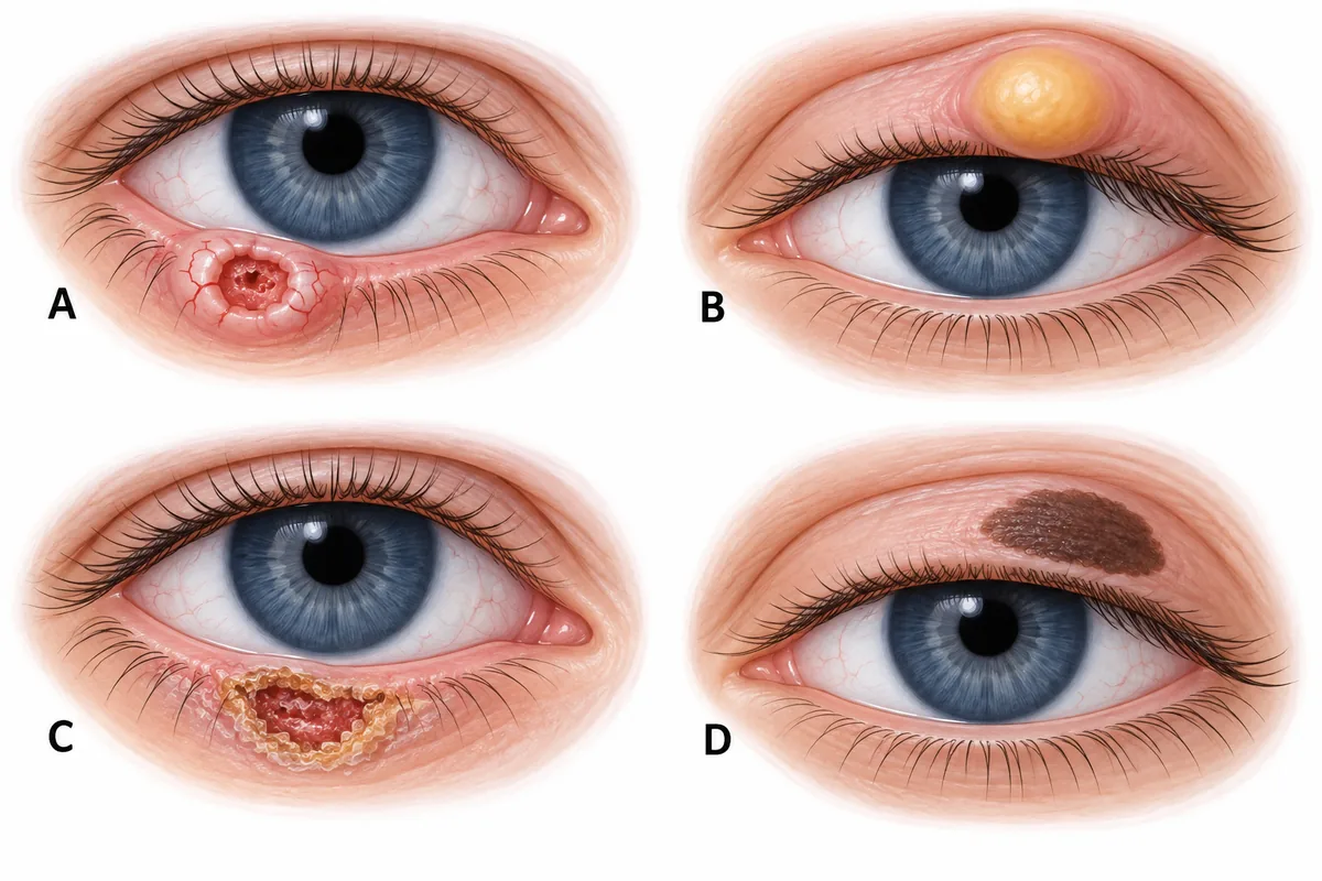
Four eyelid lesions are drawn in the figure. Which one is the commonest eyelid malignancy? Four eyelid lesions drawn at the same scale, on the lower or upper lid.
Look first: compare the edge, surface and border of each lesion before judging its size.
 View larger ↗⤢ Tap to enlarge
- ALesion A
- BLesion B
- CLesion C
- DLesion D
Show the answer and explanation
Answer: A. Lesion A
A. Lesion A. Its raised, rolled, pearly edge with fine blood vessels running across it is the classic appearance of basal cell carcinoma, the commonest malignant tumour of the eyelid. Basal cell carcinoma is locally invasive but only rarely spreads to distant sites. Read the edge first: rolled, pearly and carrying vessels.
Why the other options lose: Lesion B (B) is a firm yellow nodule that resembles a benign lid cyst. That resemblance is why sebaceous carcinoma is missed so often, but it is far less common than lesion A. Lesion C (C) is a crusted, keratotic ulcer with an irregular edge, which suggests squamous cell carcinoma instead. Lesion D (D) is flat and evenly pigmented with a regular border: a benign pigmented lesion, not a malignancy.
Recallab question 55843 · Dermatology › Skin Cancer · reference: Fitzpatrick's Dermatology
Recallab example 2 · four surfaces, one disease
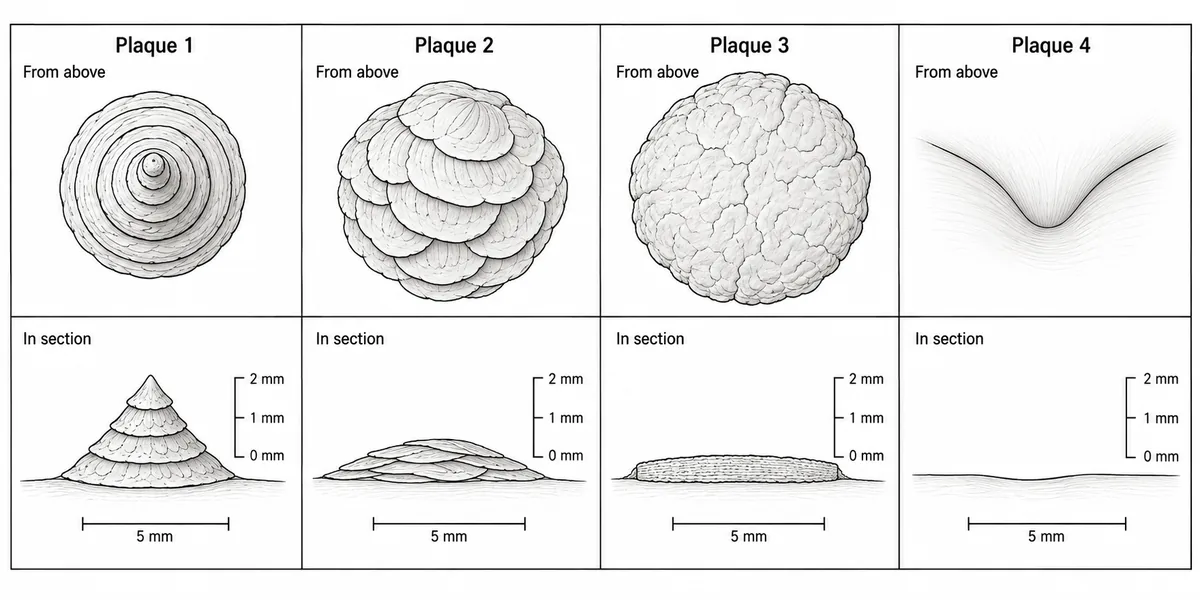
Four patients with the same chronic scaling disease, none of whom has been treated for years, have one plaque each drawn magnified in the figure, from above and in section. The diagnosis is the same in all four and is not in question. Which plaque is the ostraceous form? Four plaques of one chronic scaling disease, each drawn magnified from above and in section.
Look first: in each section, see how high the scale stands and how its layers lie.
 View larger ↗⤢ Tap to enlarge
- ABroad overlapping flat plates
- BSmooth unscaled plaque in a fold
- COne huge uniformly thick plaque
- DConcentric raised cones
Show the answer and explanation
Answer: A. Broad overlapping flat plates
A. Broad overlapping flat plates. The diagnosis is the same in all four panels; only the shape of the surface differs, and the named forms of this disease are named after that shape. An oyster shell is built of broad flat plates overlapping each other. Plaque 2 is built exactly that way in section and stays low: that is the ostraceous form.
Why the other options lose: Smooth unscaled plaque in a fold (B) is the inverse form: moisture in a fold rubs the scale away as fast as it forms, so the surface is glazed rather than built up. One huge uniformly thick plaque (C) is named for its extent and thickness rather than the shape of its surface; that is the elephantine form. Concentric raised cones (D) stand up from the skin like a limpet shell, the rupioid form. Limpet stands up; oyster lies flat.
Recallab question 59641 · Dermatology › Psoriasis · reference: Fitzpatrick's Dermatology
Recallab example 3 · one shin among four
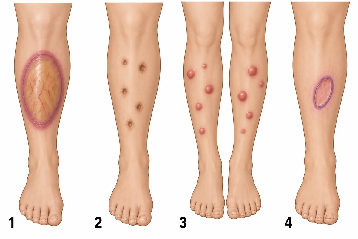
The front of the shins of a woman with type 1 diabetes is drawn in the figure as panel 1, beside three other shin lesions. What is lesion 1? A diabetic woman's shin beside three other shin lesions.
Look first: compare the colour, surface and edge of lesion 1 with the three panels beside it.
 View larger ↗⤢ Tap to enlarge
- APyoderma gangrenosum
- BDiabetic dermopathy
- CErythema nodosum
- DNecrobiosis lipoidica
Show the answer and explanation
Answer: D. Necrobiosis lipoidica
D. Necrobiosis lipoidica. Lesion 1 is a shiny, thin, yellow-brown plaque on the shin with fine vessels running across it and a slightly raised violet-red edge. That is necrobiosis lipoidica, strongly associated with diabetes; biopsy shows palisading granulomas around degenerate collagen. The plaques can appear before diabetes is diagnosed and do not track with how well the glucose is controlled.
Why the other options lose: Pyoderma gangrenosum (A) is panel 4, an ulcer with a ragged purple overhanging edge. Diabetic dermopathy (B) is panel 2: small brown depressed scars without granulomas. Erythema nodosum (C) is panel 3, tender red nodules. Yellow, shiny, thin and on the shin points to lesion 1.
Recallab question 55439 · Dermatology › Systemic Conditions · reference: Fitzpatrick's Dermatology
Read the shape, the movement and what comes with it
Recallab example 4 · a band from brow to scalp
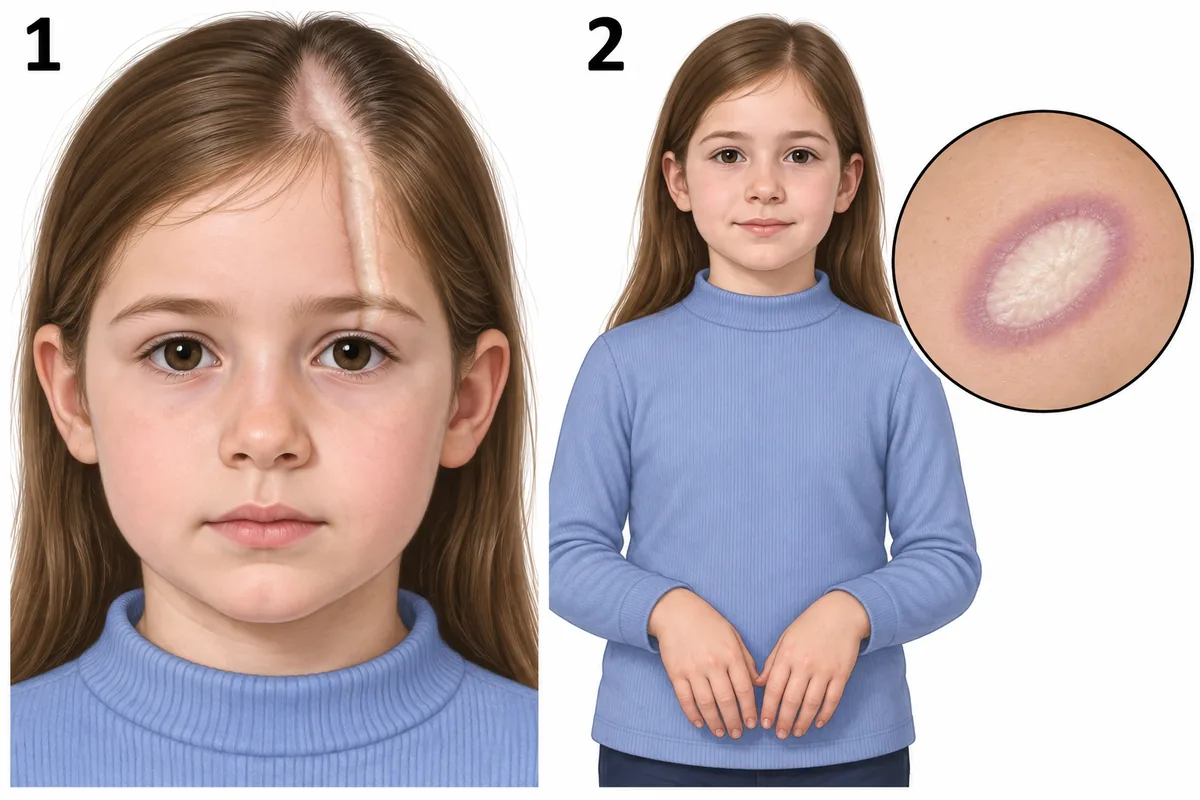
A nine-year-old girl is drawn in the figure with the skin change on her forehead and a second lesion on her trunk. What is the diagnosis? A child's face and trunk, each with a skin lesion drawn.
Look first: follow the forehead band into the hairline, then check the trunk lesion and the hands.
 View larger ↗⤢ Tap to enlarge
- ADiscoid lupus
- BLinear morphoea
- CSystemic sclerosis
- DLichen striatus
Show the answer and explanation
Answer: B. Linear morphoea
B. Linear morphoea. The forehead band is a linear, depressed, ivory groove with scarring hair loss where it crosses the scalp. That frontoparietal pattern is called en coup de sabre, the sword cut. The trunk plaque with a lilac ring is an active morphoea plaque. Morphoea is localised scleroderma of the skin and underlying tissue, and the hands are drawn normal, without the tight fingers and colour change of systemic disease.
Why the other options lose: Discoid lupus (A) forms scaly plaques with plugged follicles; it does not form an indurated linear groove. Systemic sclerosis (C) brings the Raynaud phenomenon and sclerodactyly, both absent here, and a linear band is not its pattern. Lichen striatus (D) is a transient linear papular rash of a limb, not a sclerotic groove on the forehead. Morphoea stays in the skin; systemic sclerosis starts in the fingers.
Recallab question 55639 · Dermatology › Autoimmune Conditions · reference: Fitzpatrick's Dermatology
Recallab example 5 · one back, two days
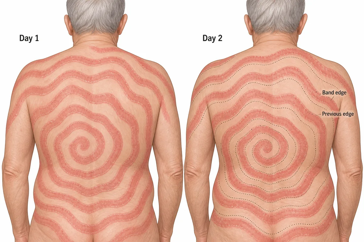
A 64-year-old man who has smoked for forty years has an itchy eruption over his trunk. His back is drawn on two consecutive days in the figure. What should be done next? The same back drawn on day 1 and day 2.
Look first: compare each band edge on day 2 with the dotted line that marks it on day 1.
 View larger ↗⤢ Tap to enlarge
- APotent topical steroid alone
- BScreen for hidden cancer
- CPatch test for allergens
- DReassure and observe
Show the answer and explanation
Answer: B. Screen for hidden cancer
B. Screen for hidden cancer. Concentric wavy bands with trailing scale give a wood-grain pattern, and the band edges move about a centimetre a day between the two drawings. That rapid migration marks erythema gyratum repens. Most cases accompany an internal malignancy, lung cancer commonest, so in a heavy smoker the chest is imaged first. The eruption may appear before, with or after the tumour is found, so a first negative search is repeated.
Why the other options lose: Potent topical steroid alone (A) treats the skin and misses the cancer; this is not an eczema. Patch test for allergens (C) suits contact dermatitis, which stays at the site of contact and does not form migrating concentric bands. Reassure and observe (D) suits erythema annulare centrifugum, which spreads slowly without this wood-grain pattern.
Recallab question 57943 · Dermatology › Dermatitis Erythema · reference: Fitzpatrick's Dermatology
Recallab example 6 · a violet nose and the chest beside it
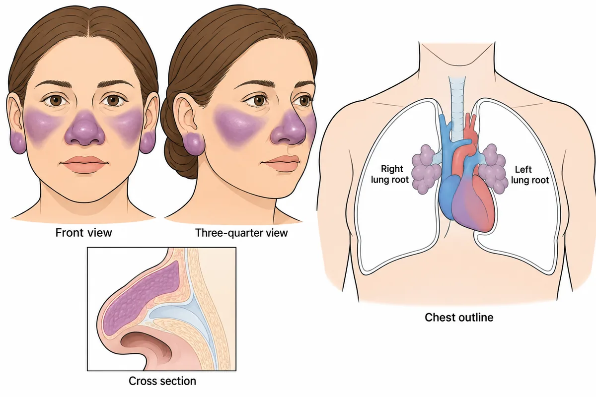
A woman of forty has had the facial change drawn in the figure for two years, and it has never been painful or itchy. The chest outline drawn beside it belongs to the same patient. What is the skin lesion? A face with a two year facial change, beside a chest outline from the same patient.
Look first: check the surface of the nose for scale or plugs, then read the lung roots in the chest outline.
 View larger ↗⤢ Tap to enlarge
- ARhinophyma of rosacea
- BDiscoid lupus erythematosus
- CNodular basal cell carcinoma
- DLupus pernio
Show the answer and explanation
Answer: D. Lupus pernio
D. Lupus pernio. The nose, cheeks and ears are indurated, shiny and dusky violet, and the cross section places the thickening in the dermis beneath an intact surface, with no scale, follicular plug or pustule. Both lung roots are enlarged and lobulated with clear lung fields beyond them. Skin and chest together point to one chronic granulomatous disorder: lupus pernio is the skin marker of chronic sarcoidosis and predicts a fibrotic course, so systemic rather than topical treatment is planned.
Why the other options lose: Rhinophyma of rosacea (A) is red and greasy with enlarged sebaceous openings, and it does not enlarge the lung roots. Discoid lupus erythematosus (B) scales, plugs follicles and leaves a depressed scarred centre. Nodular basal cell carcinoma (C) is a single pearly nodule with a rolled edge and surface vessels. A nose that is infiltrated and violet, not greasy and red, is a warning about the chest.
Recallab question 58242 · Dermatology › Systemic Conditions · reference: Fitzpatrick's Dermatology
The one-minute routine
| If the figure gives | Ask first | It settles |
|---|---|---|
| Four lesions on the eyelid | Which edge is rolled and pearly, with vessels across it? | Basal cell carcinoma, the commonest eyelid malignancy |
| Plaques of one scaling disease | Does the scale stand up as a cone or lie in flat overlapping plates? | Rupioid (limpet) or ostraceous (oyster) |
| A shin plaque in a patient with diabetes | Is it yellow, shiny and thin, with vessels across it? | Necrobiosis lipoidica |
| A depressed band from brow to scalp | Are the hands normal? | Linear morphoea (en coup de sabre), not systemic sclerosis |
| Wavy bands drawn on two days | Have the band edges moved? | Erythema gyratum repens: search for a cancer |
| A swollen violet nose | Is it infiltrated without scale, and are the lung roots enlarged? | Lupus pernio, a marker of chronic sarcoidosis |
Practise dermatology questions and work through six more skin pictures in dermatology lesions and signs.
Why candidates use Recallab
- Built towards 22,000 — a batch at a time
- Illustrations drawn for the question
- Every option explained, not just the right one
- AI study tools that are live today
- Start free, today
Or see the plans, or try a timed mock section.
Register to practise NEET PG dermatology questionsFree account, no card needed. Practise with every option explained and a reference on every question.Drawn from questions #55843, #59641, #55439, #55639, #57943, #58242 in the Recallab bank, each with its own verified answer and current-edition reference. Last reviewed 20 October 2026.