
Recallab › Blog › Dermatology for NEET PG: six skin pictures and the one thing to look at first
Dermatology for NEET PG: six skin pictures and the one thing to look at first
A skin picture is a lot of detail and one clue. The skill is to know which feature to look at first, and what it tells you.
Register to practise NEET PG dermatology questions →Free account, no card needed.
Dermatology questions show a nail, a patch or a scraping and ask what it is, or what to do about it. The pictures below are illustrations drawn for the questions, not photographs of patients. Each is a real Recallab MentorQ, and each answer turns on one feature of the drawing. Try each before you open the answer.
Before you name a skin picture: three questions
- Where is the change? On the surface, at one level of the skin, or in a structure such as the nail.
- What is the pattern? Round, ringed, cracked, scattered or confluent, and where on the body.
- What does the pattern make you do? Reassure, treat, scrape and stain, or move the patient to a higher level of care.
Nails and blisters
Recallab example 1 · the nail
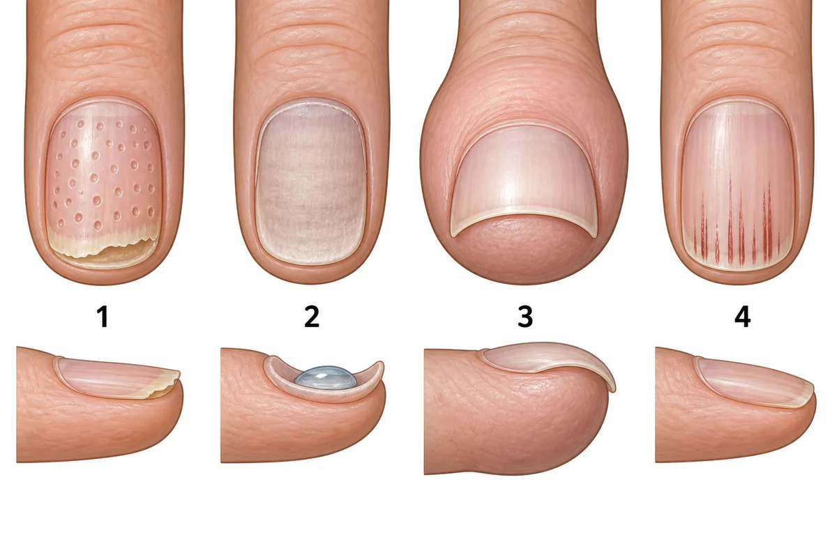
Four fingernails are drawn in the figure, each showing one change. Which nail belongs to a patient with iron deficiency?
Look first: look at each nail from the side as well as from above; the shape of the plate is the clue.
 View larger ↗⤢ Tap to enlarge
- ANail 1
- BNail 2
- CNail 4
- DNail 3
Show the answer and explanation
Answer: B. Nail 2
Nail 2, koilonychia. In profile the plate is turned up at its edges with a hollow in the middle, deep enough to hold a drop of water, which is the bedside demonstration of the sign. Iron is needed for the keratin of the plate, so deficiency makes it thin and soft. The change takes months to appear and months to reverse after treatment.
Why the closest rival loses: nail 1 (A) has pitting and a lifted free edge, which is psoriasis of the nail. Nail 3 (D) is clubbing, which points to suppurative lung disease, cyanotic heart disease or malignancy. Nail 4 (C) shows splinter haemorrhages, which raise infective endocarditis, though trauma is far commoner. Spoon for iron, drumstick for the lung.
Recallab question 55040 · Skin › Appendages, Tumours & Miscellaneous · reference: Fitzpatrick's Dermatology
Recallab example 2 · the blister
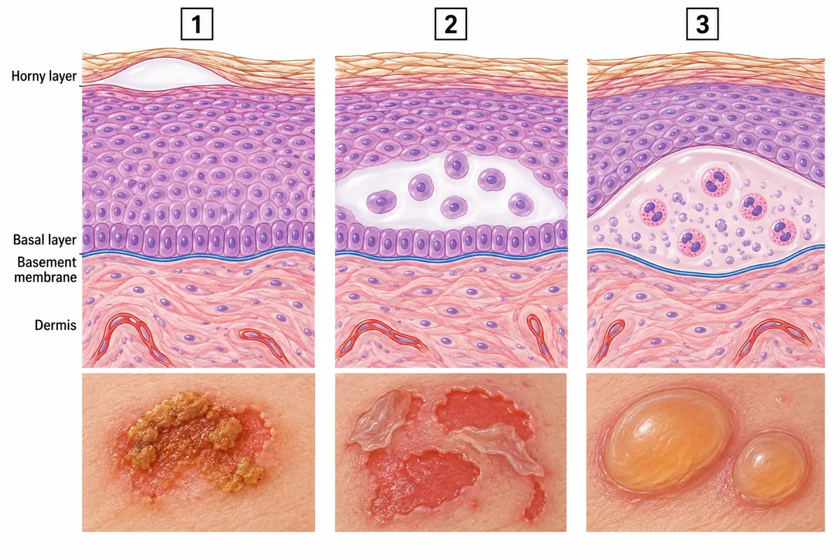
Three blisters are drawn in the figure as sections through the skin, each split at a different level. Which disease produces blister 2?
Look first: find the basement membrane first, then ask which side of it the split lies on.
 View larger ↗⤢ Tap to enlarge
- ABullous impetigo
- BDermatitis herpetiformis
- CPemphigus vulgaris
- DBullous pemphigoid
Show the answer and explanation
Answer: C. Pemphigus vulgaris
Pemphigus vulgaris. The level of the split decides how the blister behaves. In section 2 the split lies within the epidermis, just above the basal layer, which stays attached to the basement membrane. The cells above have lost their attachments to each other and float free. A roof of live epidermal cells has no strength, so the blister is flaccid and collapses early: patients present with erosions rather than intact blisters.
Why the closest rival loses: bullous pemphigoid (D) is section 3, split below the basement membrane, which is why those blisters stay tense. Bullous impetigo (A) is section 1, split just under the horny layer, the most superficial of the three. Dermatitis herpetiformis (B) also splits below the membrane. Deep split, tense blister; shallow split, raw erosion.
Recallab question 55038 · Skin › Vesiculobullous & Connective Tissue · reference: Fitzpatrick's Dermatology
Patterns you can name
Recallab example 3 · the mole
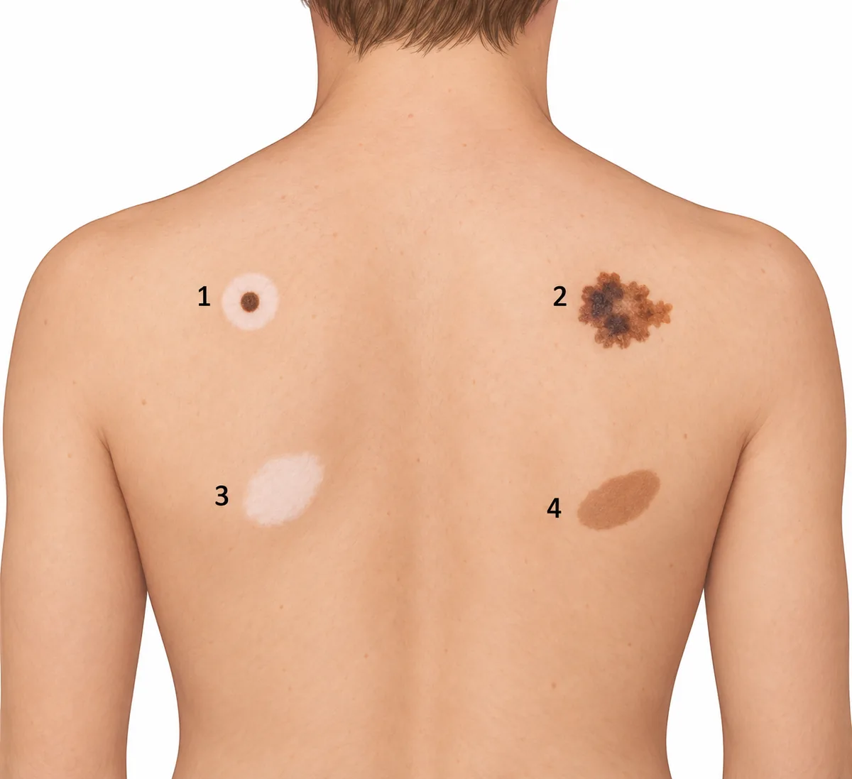
Four lesions on the back of a fifteen-year-old are drawn in the figure. What is lesion 1?
Look first: compare the four for size, colour and edge, and ask which one is regular.
 View larger ↗⤢ Tap to enlarge
- AA halo naevus
- BA patch of vitiligo
- CA Meyerson naevus
- DA regressing melanoma
Show the answer and explanation
Answer: A. A halo naevus
A halo naevus. Lesion 1 is small, even in colour and regular, sitting centrally in a symmetrical ring of white skin. Cytotoxic T cells attack the naevus cells and the melanocytes around them, which leaves a clean halo; later the mole itself may vanish. In a teenager it is benign, though the rest of the skin is examined, and it is commoner in people with vitiligo.
Why the closest rival loses: a patch of vitiligo (B) is lesion 3, a white patch with nothing in its centre. A Meyerson naevus (C) carries a red, scaly eczematous halo, not a smooth white ring. A regressing melanoma (D) is patchy and irregular; lesion 2, varied in colour with a notched border, is the worrying one.
Recallab question 55238 · Skin › Disorder Of Pigmentation · reference: Fitzpatrick's Dermatology
Recallab example 4 · the shins
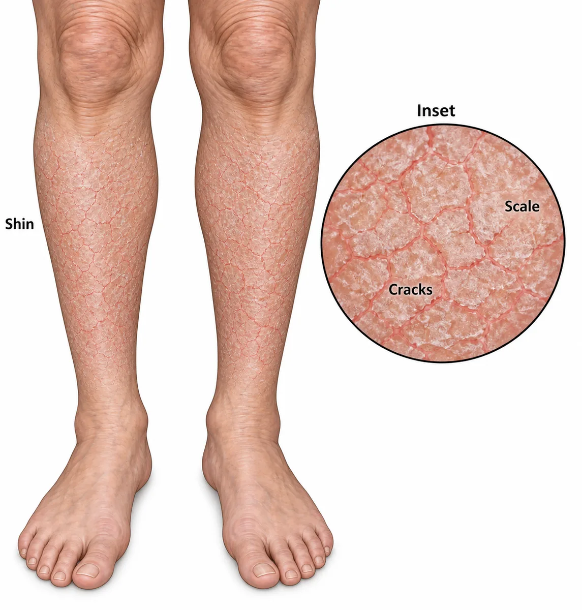
An 80-year-old man has had itchy legs all winter. He bathes twice a day in hot water with soap. Both shins are drawn in the figure. What is the diagnosis?
Look first: read the surface of the skin as a whole, then ask what the history has done to it.
 View larger ↗⤢ Tap to enlarge
- ADiscoid eczema
- BAsteatotic eczema
- CIchthyosis vulgaris
- DVenous eczema
Show the answer and explanation
Answer: B. Asteatotic eczema
Asteatotic eczema. Older skin makes less surface lipid and loses water easily, and winter air, hot water and soap strip what barrier is left. The dry, scaly skin develops a network of fine red fissures that divides the surface into polygons, like crazy paving, and the shins are the commonest site. Soap substitutes and heavy emollients restore the barrier.
Why the closest rival loses: discoid eczema (A) forms separate, coin-shaped, often weeping plaques, not a continuous cracked network. Venous eczema (D) comes with ankle swelling, brown staining and varicose veins, none of which is in the story. Ichthyosis vulgaris (C) is lifelong, with fine scale, and does not appear for the first time in old age.
Recallab question 57942 · Skin › Eczema · reference: Fitzpatrick's Dermatology
Beyond the surface
Recallab example 5 · the scraping
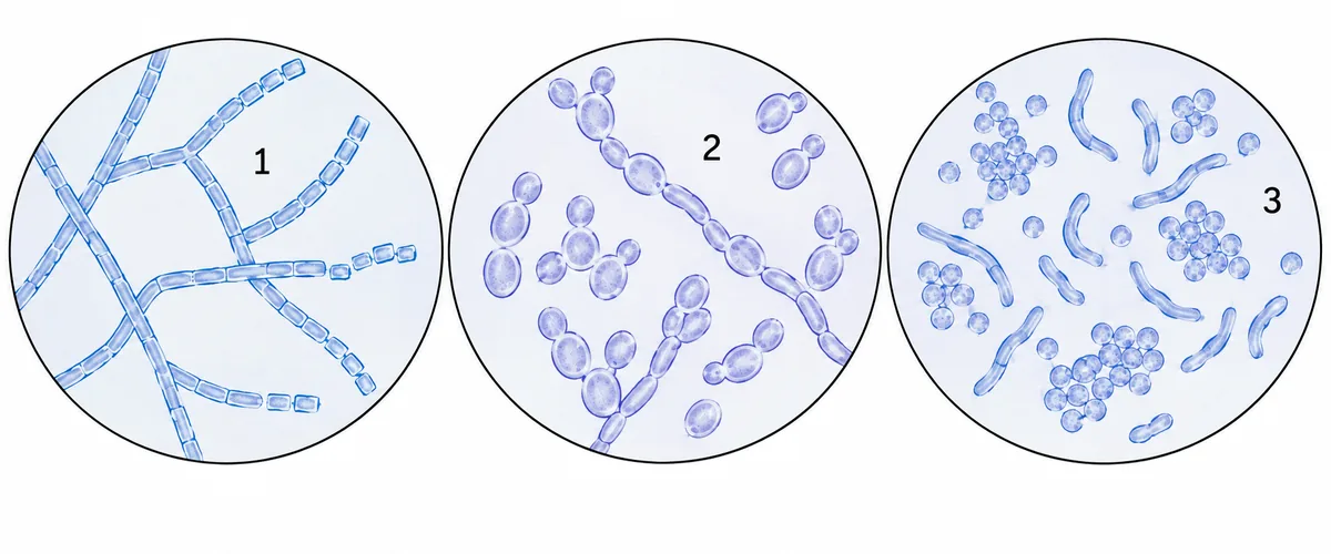
Three potassium hydroxide mounts are drawn in the figure. An itchy annular scaly plaque on the thigh has a raised active edge and central clearing. Which numbered mount matches it?
Look first: match the plaque to a diagnosis first, then ask what that organism looks like under the microscope.
 View larger ↗⤢ Tap to enlarge
- AMount 1, dermatophyte hyphae
- BMount 3, Malassezia
- CMount 3, dermatophyte hyphae
- DMount 2, Candida
Show the answer and explanation
Answer: A. Mount 1, dermatophyte hyphae
Mount 1, dermatophyte hyphae. An annular plaque with a raised, active edge and central clearing is classic tinea corporis. Dermatophytes show as long, branching, septate hyphae that break into chains of arthrospores, as in mount 1. Misusing a topical steroid on an unrecognised dermatophyte infection produces tinea incognito, where the active edge is lost and the diagnosis gets harder.
Why the closest rival loses: mount 3 shows short, curved hyphae with clusters of round spores, the appearance of Malassezia, not the long branching septate hyphae of a dermatophyte. So option C names the wrong mount for a dermatophyte, and option B names the right organism for mount 3 but the wrong disease: Malassezia causes patchy pigment change, not a spreading annular edge. Mount 2 (D) shows budding yeast and pseudohyphae, which fits Candida in moist folds with satellite pustules, not a plaque on the thigh.
Recallab question 55358 · Skin › Fungal Infection · reference: Fitzpatrick's Dermatology
Recallab example 6 · the drug reaction
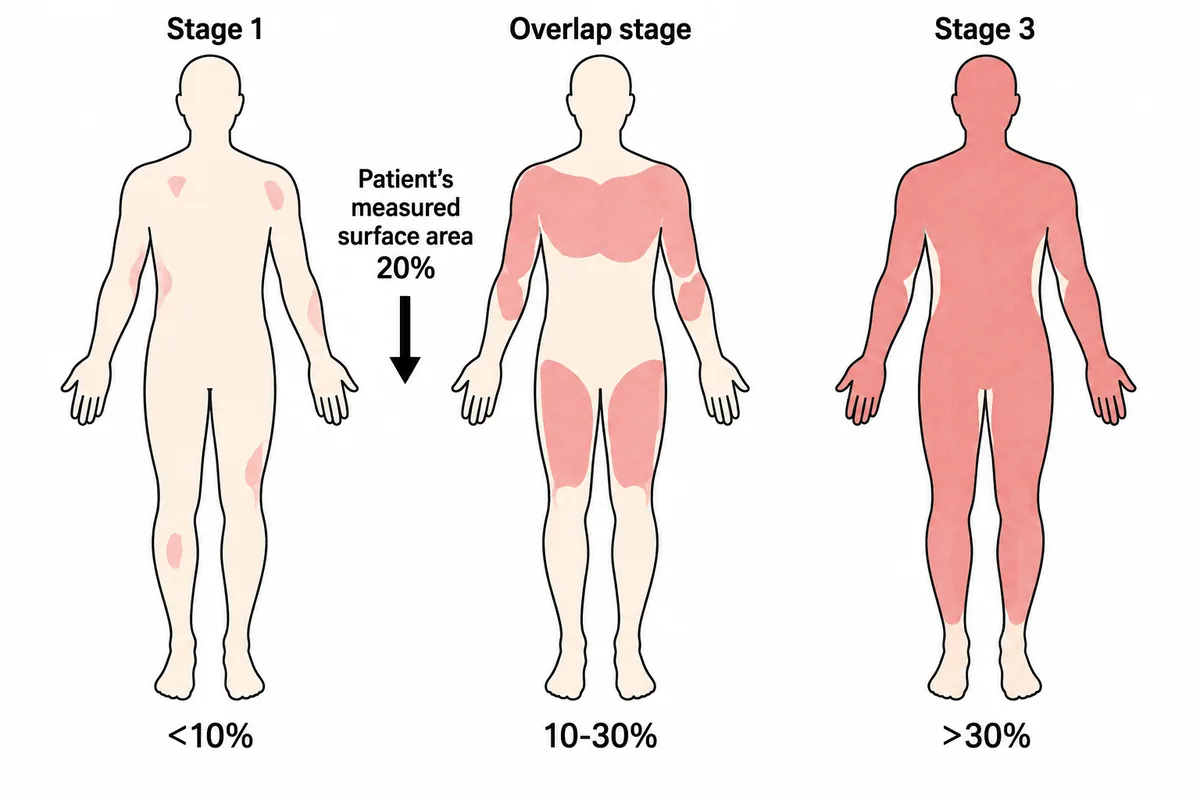
Three body outlines are drawn in the figure, each with the percentage of detached skin that marks one stage of the same severe drug reaction, and one patient's measured detachment of 20 per cent is marked. The drug has already been stopped at the district hospital. Where should she now be cared for?
Look first: read the percentage on the scale, then ask what the lost skin cannot do any more.
 View larger ↗⤢ Tap to enlarge
- AA burns or intensive care unit
- BDischarge with outpatient review
- CA general medical ward
- DAn isolation ward
Show the answer and explanation
Answer: A. A burns or intensive care unit
A burns or intensive care unit. The outlines are staged by the share of skin that has detached: under 10 per cent, 10 to 30 per cent, over 30 per cent. At 20 per cent she sits in the overlap band between Stevens-Johnson syndrome and toxic epidermal necrolysis. Detached epidermis loses fluid, protein and heat and lets infection in, as a burn does, so she needs warming, barrier nursing, fluid balance and wound care of burns-unit standard, and the risk grows with the area lost.
Why the closest rival loses: a general medical ward (C) cannot give the fluid, temperature and wound care that a burn of this size needs. Discharge (B) is out of the question: a fifth of the skin detached is life-threatening. An isolation ward (D) answers an infection: staphylococcal scalded skin syndrome follows one and splits the skin high in the epidermis. This is a drug reaction. Under 10 per cent is Stevens-Johnson, over 30 is toxic epidermal necrolysis, and the culprit drug is stopped at once.
Recallab question 56242 · Skin › Drugs & Drug reactions · reference: Fitzpatrick's Dermatology
The one-minute routine
| If you see | Ask first | It usually settles |
|---|---|---|
| Four fingernails | What shape is the plate from the side | Which systemic cause |
| Blisters drawn as sections | Which side of the basement membrane is the split | Flaccid or tense, and which disease |
| Lesions on a back | Which is small, even and regular | Benign or worrying |
| Shins of an older person | What has the history done to the skin | Eczema type, and the treatment |
| A scraping | What does the plaque make you suspect | Which organism shows in the mount |
| Body outlines with percentages | Where does the patient's value fall | The level of care |
For other kinds of figure, see ten NEET PG image questions and six eye and ENT spotters. For the decision that follows the picture, see next-best-step management questions.
For pregnancy and the antenatal visit, see six NEET PG antenatal care questions.
Why candidates use Recallab
- Built towards 22,000 — a batch at a time
- Illustrations drawn for the question
- Every option explained, not just the right one
- AI study tools that are live today
- Start free, today
Or see the plans, or try a timed mock section.
Register to practise NEET PG dermatology questionsFree account, no card needed. Practise with every option explained and a reference on every question.Drawn from questions #55040, #55038, #55238, #57942, #55358, #56242 in the Recallab bank, each with its own verified answer and current-edition reference. Last reviewed 7 October 2026.