
Recallab › Blog › Read the figure first: ten NEET PG image questions and what to look at in each
Read the figure first: ten NEET PG image questions and what to look at in each
An image question rewards the candidate who describes before naming. Here are ten, each with a different kind of figure, and the one thing to look at before you read the options.
Register to practise NEET PG image MCQs →Free account, no card needed.
NEET PG pictures go well beyond X-rays: traces, scans, eyes, skin, stains, specimens and bedside tests all appear. Each asks you to see one feature clearly and connect it to the question. The ten below are real Recallab MentorQs, shown with the figure the learner sees. Try each one, then open the answer.
Three questions to ask any figure
- What kind of image is this? A trace, a scan, a stain, a specimen, a bedside test. The kind of image tells you what can and cannot be shown.
- What is the one abnormal feature, or the one thing that differs between panels? Say it in plain words before you reach for a diagnosis.
- What does the question actually ask about it? A diagnosis, an organism, a mechanism or the next step. Recognising the picture is not the same as answering the question.
That last step is the subject of yesterday’s post on question types. Here the emphasis is on the first two.
Traces and scans
Recallab example 1 · heart-sound trace
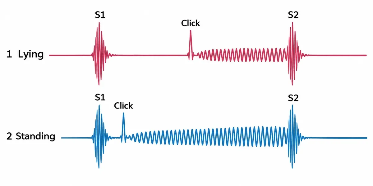
Phonocardiograms of a young woman with palpitations are drawn in the figure, lying and then standing. What is the diagnosis?
Look first: line the two traces up. Find the click in each, and ask where it sits between S1 and S2 when she stands.
 View larger ↗⤢ Tap to enlarge
- AHypertrophic cardiomyopathy
- BAortic stenosis
- CMitral valve prolapse
- DAtrial septal defect
Show the answer and explanation
Answer: C. Mitral valve prolapse
Decisive feature: a mid-systolic click followed by a late systolic murmur, and on standing both move towards S1. A smaller ventricle lets the leaflets prolapse sooner, so the click comes earlier and the murmur lengthens. Squatting does the opposite.
Why the closest rival loses: the murmur of hypertrophic cardiomyopathy (A) also grows on standing, but there is no click. The ejection click of aortic stenosis (B) sits just after S1 and does not move with posture; an atrial septal defect (D) gives a fixed split second sound and no click.
Recallab question 55272 · Medicine › Cardiology · reference: Harrison's Principles of Internal Medicine
Recallab example 2 · ventilation–perfusion scan
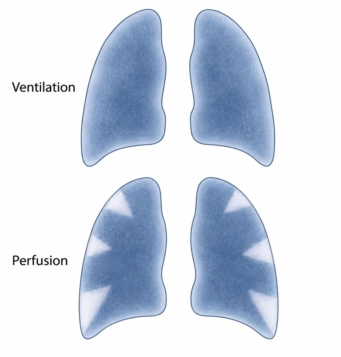
Paired ventilation and perfusion lung scans are drawn in the figure. A woman is breathless on the eighth day after a hip replacement, and her chest radiograph is normal. What is the diagnosis?
Look first: compare the two scans lung by lung. Where does air reach, and where does blood not?
 View larger ↗⤢ Tap to enlarge
- APneumonia with effusion
- BPulmonary embolism
- CA normal study
- DChronic obstructive pulmonary disease
Show the answer and explanation
Answer: B. Pulmonary embolism
Decisive feature: ventilation is even throughout, but perfusion has wedge-shaped gaps: a mismatch. Blood flow is blocked where air still arrives. With breathlessness after a hip replacement and a normal radiograph, that is a high probability of pulmonary embolism.
Why the closest rival loses: pneumonia (A) gives a single matched defect, often beside an effusion. Chronic obstructive pulmonary disease (D) gives matched defects, with ventilation missing too. The wedges rule out a normal study (C).
Recallab question 55353 · Medicine › Respiratory · reference: Harrison's Principles of Internal Medicine
Recallab example 3 · serial chest films
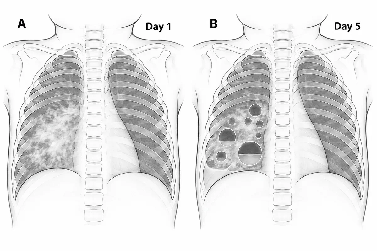
Two chest films taken five days apart in a febrile, breathless eight-month-old are drawn in the figure. Which organism most likely caused the pneumonia?
Look first: put day 1 beside day 5. What has appeared in the right lung that was not there before, and what are its walls like?
 View larger ↗⤢ Tap to enlarge
- AMycobacterium tuberculosis
- BKlebsiella pneumoniae
- CStreptococcus pneumoniae
- DStaphylococcus aureus
Show the answer and explanation
Answer: D. Staphylococcus aureus
Decisive feature: thin-walled air cysts that were not there on day 1. These are pneumatoceles, and appearing within days during an infant's pneumonia they point to Staphylococcus aureus. Most shrink over weeks with treatment, but a tense one can rupture into a pneumothorax.
Why the closest rival loses: pneumococcal pneumonia (C) gives lobar consolidation and rarely forms pneumatoceles. Klebsiella (B) gives a lobar consolidation in debilitated adults; primary tuberculosis in infants (A) gives hilar nodes, not thin-walled cysts within days.
Recallab question 55650 · Paediatrics › Respiratory · reference: Ghai Essential Pediatrics
Recallab example 4 · MRI
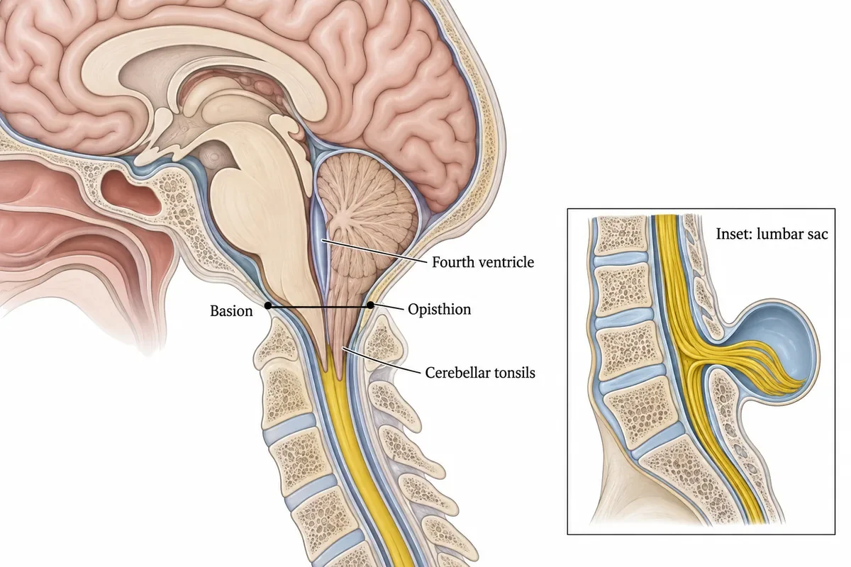
A midsagittal MRI of the posterior fossa is drawn in the figure, with a line drawn from the front to the back of the foramen magnum and the cerebellar tonsils traced below it. An inset shows a lumbar swelling present since birth. What is the diagnosis?
Look first: follow the line across the foramen magnum and ask how far below it the tonsils reach, then what the lumbar inset adds.
 View larger ↗⤢ Tap to enlarge
- AChiari II malformation
- BChiari I malformation
- CDandy-Walker malformation
- DPlatybasia
Show the answer and explanation
Answer: A. Chiari II malformation
Decisive feature: peg-like tonsils well below the basion–opisthion line in a small posterior fossa, together with a lumbar myelomeningocele. That pairing is Chiari II. Crowding at the foramen magnum often obstructs the flow of cerebrospinal fluid and causes hydrocephalus.
Why the closest rival loses: Chiari I (B) shows tonsillar descent of a few millimetres in a normal-sized posterior fossa, usually with no spinal defect. Dandy–Walker (C) is a cystic, enlarged fourth ventricle with an absent vermis; platybasia (D) is a flattened skull base that does not explain the spinal lesion.
Recallab question 55682 · Radiology › Neuroimaging, head and neck · reference: Harrison's Principles of Internal Medicine
At the bedside
Recallab example 5 · slit lamp
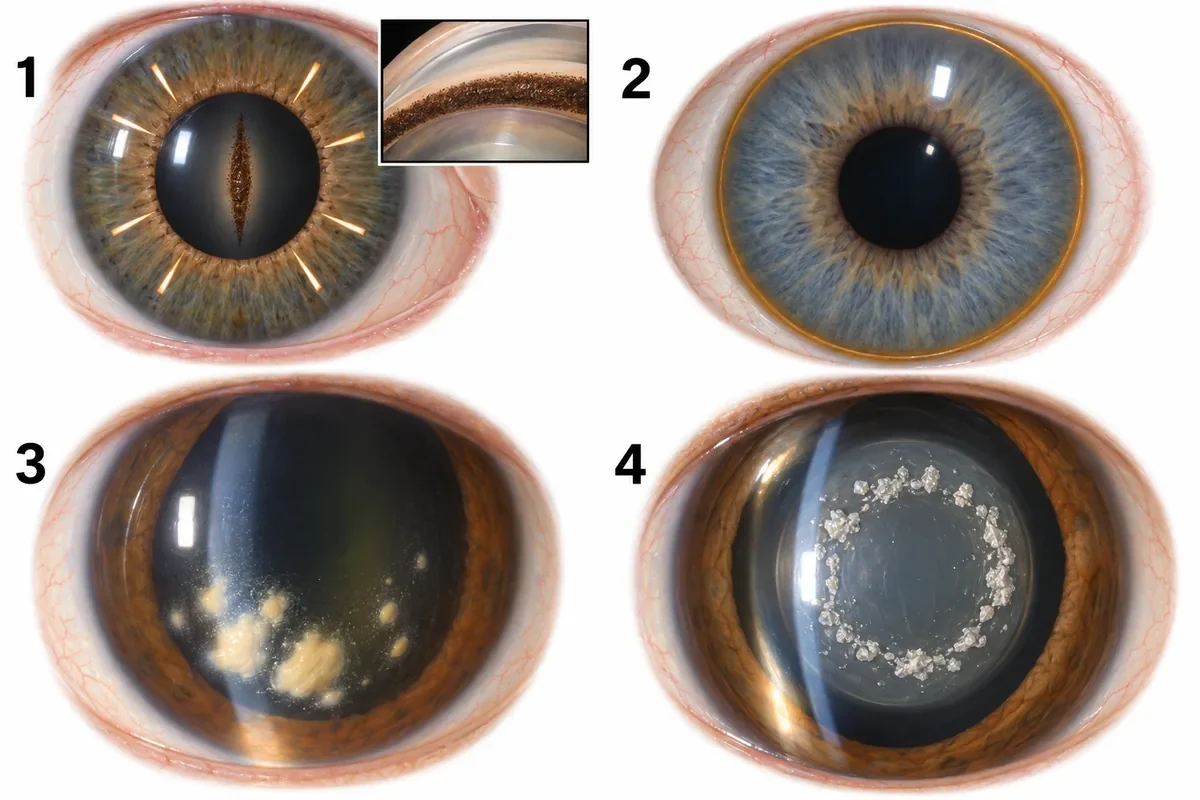
The front of the eye of a young myopic man is drawn in the figure as panel 1, beside three other slit-lamp appearances. What is the diagnosis?
Look first: panel 1 is the patient; the other three are there to be ruled out. Look at the cornea, the iris and the inset of the drainage angle.
 View larger ↗⤢ Tap to enlarge
- APigment dispersion syndrome
- BPseudoexfoliation syndrome
- CGranulomatous uveitis
- DWilson disease
Show the answer and explanation
Answer: A. Pigment dispersion syndrome
Decisive feature: a vertical spindle of pigment on the corneal endothelium (a Krukenberg spindle), spoke-like transillumination defects in the mid-peripheral iris and a heavily pigmented angle, in a young myopic man. That is pigment dispersion syndrome, which can lead to pigmentary glaucoma.
Why the closest rival loses: pseudoexfoliation (B) lays white flakes on the lens (panel 4), in older people. Panel 3 shows the mutton-fat keratic precipitates of granulomatous uveitis (C), and panel 2 the Kayser–Fleischer ring of Wilson disease (D).
Recallab question 55487 · Ophthalmology › Glaucoma · reference: Khurana, Comprehensive Ophthalmology
Recallab example 6 · skin and smear
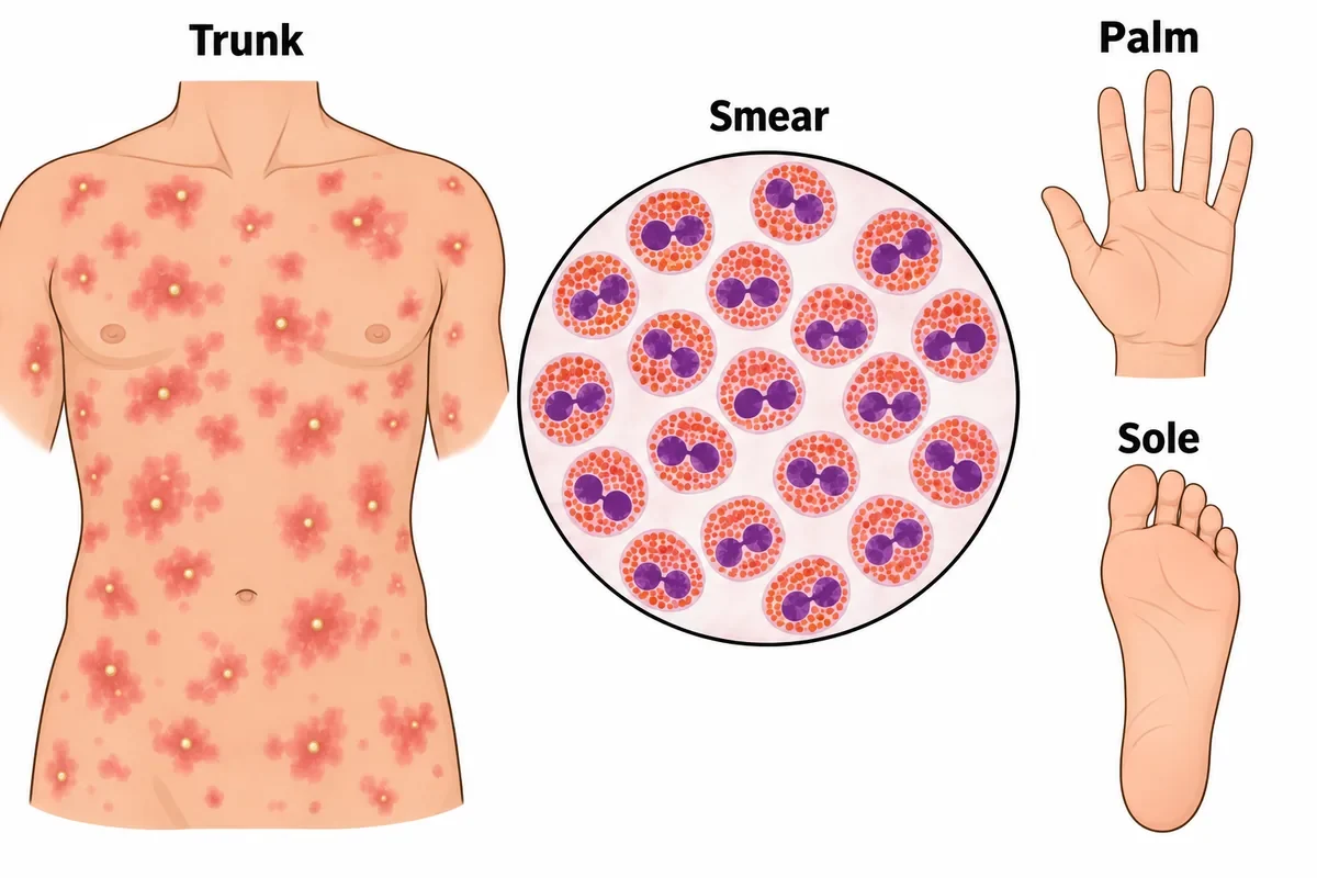
The trunk of a well two-day-old baby is drawn in the figure, with a stained smear of the contents of one pustule beside it. What is the rash?
Look first: two clues. Where the rash is and is not (check the palms and soles), and which cell fills the smear.
 View larger ↗⤢ Tap to enlarge
- ANeonatal herpes simplex
- BStaphylococcal pustulosis
- CErythema toxicum neonatorum
- DTransient neonatal pustular melanosis
Show the answer and explanation
Answer: C. Erythema toxicum neonatorum
Decisive feature: red blotches with central pustules on day two, palms and soles spared, and a smear full of eosinophils (two-lobed nuclei, orange-red granules) with no bacteria. That is erythema toxicum neonatorum: sterile, self-limiting, and treated by reassuring the parents.
Why the closest rival loses: staphylococcal pustules (B) would show neutrophils and Gram-positive cocci. Herpes (A) gives clustered vesicles in an unwell baby; transient neonatal pustular melanosis (D) is present at birth, often involves palms and soles, contains neutrophils and leaves pigmented macules.
Recallab question 55248 · Paediatrics › Newborn infants · reference: Ghai Essential Pediatrics
Under the microscope and on the cut surface
Recallab example 7 · immunostains
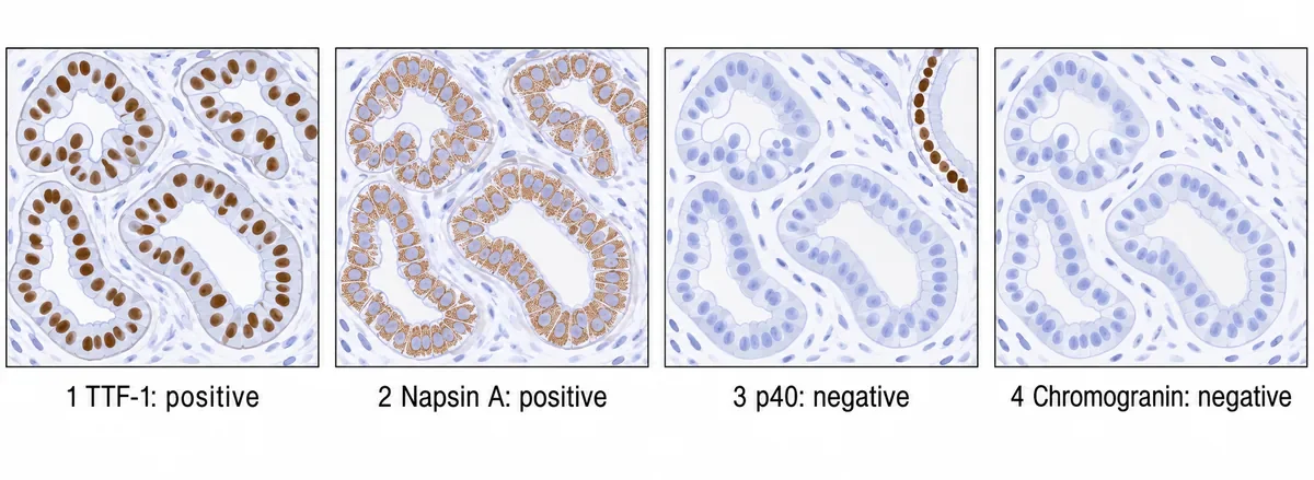
A peripheral lung mass in a woman who has never smoked is biopsied. Four immunostains on the tumour are drawn in the figure, each with its result. Which tumour is it?
Look first: read the four results as one sentence, and check that the negative stain has something positive in the field to prove it worked.
 View larger ↗⤢ Tap to enlarge
- ALarge cell neuroendocrine carcinoma
- BAdenocarcinoma
- CSquamous cell carcinoma
- DSmall cell carcinoma
Show the answer and explanation
Answer: B. Adenocarcinoma
Decisive feature: TTF-1 (nuclear) and napsin A (cytoplasmic) are positive, so the tumour is a lung adenocarcinoma. p40 is negative, and the brown basal cells at the edge of panel 3 show the stain itself worked. Chromogranin is negative, so a neuroendocrine tumour is unlikely.
Why the closest rival loses: squamous carcinoma (C) would be p40 positive. Small cell carcinoma (D) can be TTF-1 positive, but it is napsin A negative and positive for neuroendocrine markers, as large cell neuroendocrine carcinoma (A) would be.
Recallab question 55717 · Pathology › Respiratory · reference: Robbins, Cotran & Kumar Pathologic Basis of Disease
Recallab example 8 · gross specimens
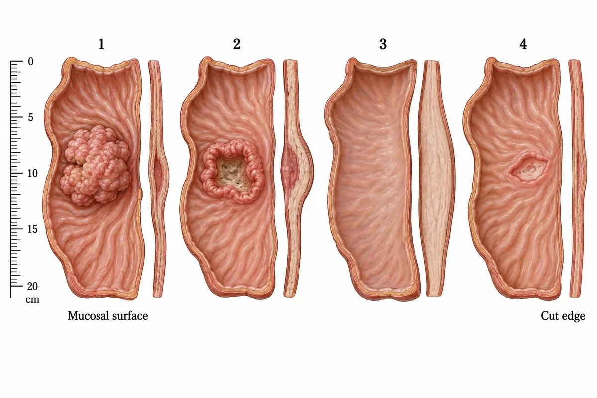
Four resected segments of stomach are drawn in the figure, opened along the greater curvature. Which specimen shows the pattern that carries the worst outlook?
Look first: look at the cut edge beside each specimen, not only the mucosal surface.
 View larger ↗⤢ Tap to enlarge
- ASpecimen 2
- BSpecimen 1
- CSpecimen 4
- DSpecimen 3
Show the answer and explanation
Answer: D. Specimen 3
Decisive feature: specimen 3 has a thick, rigid wall under almost normal mucosa. That is linitis plastica: the tumour spreads diffusely within the wall and provokes fibrosis instead of growing into the lumen. Superficial biopsies can come back negative, so the diagnosis is late and spread is usual by then.
Why the closest rival loses: the ulcer with rolled, everted margins (specimen 2) is malignant but localised. The polypoid mass (specimen 1) is easily seen and biopsied; the flat ulcer with a clean base (specimen 4) looks benign.
Recallab question 55059 · Pathology › Neoplasia · reference: Robbins, Cotran & Kumar Pathologic Basis of Disease
In the side room
Recallab example 9 · urine dipstick
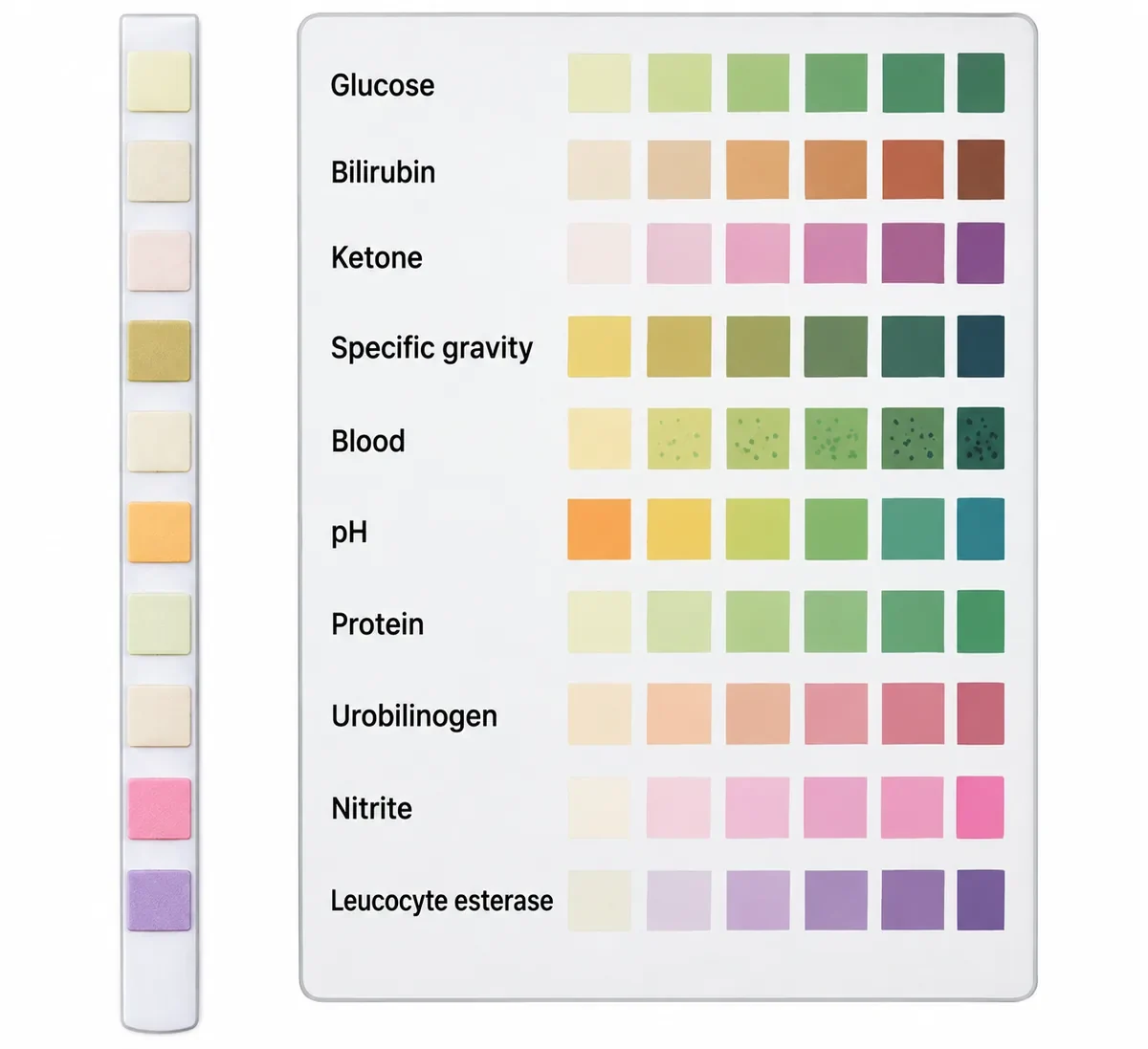
A dipstick is drawn in the figure beside its colour chart. Two pads have changed and the rest have not. Which single finding does that combination point to, given that the blood pad is unchanged?
Look first: match each pad to its row on the chart, and list only the pads that have moved.
 View larger ↗⤢ Tap to enlarge
- ASample contamination
- BBacteriuria with pyuria
- CDiabetic ketoacidosis
- DGlomerular bleeding
Show the answer and explanation
Answer: B. Bacteriuria with pyuria
Decisive feature: the nitrite and leucocyte esterase pads have changed. Nitrite means bacteria have reduced nitrate in urine held in the bladder; leucocyte esterase comes from neutrophils. Together they mean infection with pyuria. Nitrite is specific but insensitive, so a negative pad does not exclude infection.
Why the closest rival loses: contamination (A) cannot produce nitrite, which needs urine to sit in the bladder for hours. The glucose and ketone pads are unchanged, which rules out diabetic ketoacidosis (C); the unchanged blood pad rules out glomerular bleeding (D).
Recallab question 55007 · Physiology › Renal and acid–base · reference: Ganong's Review of Medical Physiology
Recallab example 10 · blood grouping
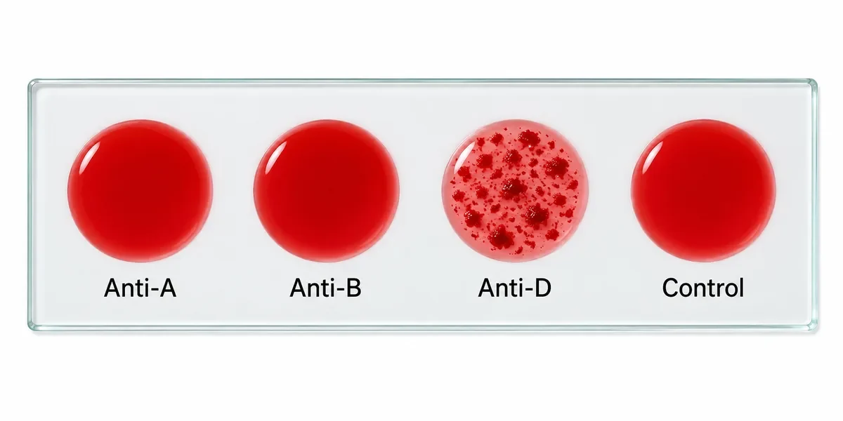
A slide grouping test is drawn in the figure, with four drops of the patient's blood each mixed with a reagent. What is the patient's blood group?
Look first: check the control first, then read each drop as present (clumped) or absent (smooth).
 View larger ↗⤢ Tap to enlarge
- AO RhD positive
- BAB RhD positive
- CO RhD negative
- DA RhD positive
Show the answer and explanation
Answer: A. O RhD positive
Decisive feature: no clumping with anti-A or anti-B, so the cells carry neither antigen: group O. Clumping with anti-D means RhD positive, and the smooth control makes the test valid.
Why the closest rival loses: RhD-negative cells (C) would not clump with anti-D. Group A (D) would clump with anti-A, and AB (B) with both. In practice, this forward grouping is always checked against reverse grouping before blood is issued.
Recallab question 55453 · Pathology › Transfusion medicine · reference: Harrison's Principles of Internal Medicine
Make it a habit
Ten figures, one routine: name the kind of image, say the one abnormal feature in plain words, then answer the question actually asked. Practise it with the explanation closed, and come back to the same figure a few days later to check you still see what matters.
If a figure still puzzles you after the explanation, the Professor in each MentorQ reads the question’s figure as well as its text; see how MentorQs work. For why a well-drawn figure helps you remember, read why illustrated MCQs belong in your NEET PG preparation.
Why candidates use Recallab
- Built towards 22,000 — a batch at a time
- Illustrations drawn for the question
- Every option explained, not just the right one
- AI study tools that are live today
- Start free, today
Or see the plans, or try a timed mock section.
Register to practise NEET PG image MCQsFree account, no card needed. Practise with every option explained and a reference on every question.Drawn from questions #55272, #55353, #55650, #55682, #55487, #55248, #55717, #55059, #55007, #55453 in the Recallab bank, each with its own verified answer and current-edition reference. Last reviewed 23 September 2026.