
Recallab › Blog › Why illustrated MCQs belong in your NEET PG preparation
Why illustrated MCQs belong in your NEET PG preparation
See the detail. Understand the relationship. Practise remembering it.
Register to practise NEET PG image MCQs →Free account, no card needed.
You answer an MCQ, read the explanation and think, “Yes, I know this.” A few weeks later, a similar question appears—and the detail you need has disappeared.
That gap between understanding something today and recalling it on exam day is one of the hardest parts of NEET PG preparation. A well-designed illustration can help bridge it. A magnified detail can make a mechanism easier to follow. An anatomical diagram can turn a list of structures into a relationship you can picture. A comparison table can make two confusing diagnoses easier to separate.
The strongest approach brings these visuals together with questions, explanations and repeated recall. Here are four examples of illustrated MCQs from Recallab’s own illustration library—and how to use them.
Image-based questions and illustrated explanations do different jobs
An image in the question asks you to observe and interpret: identify a structure, recognise a pattern or find the feature that changes the answer. It should give you the evidence needed to reason without labelling the answer for you.
An image in the explanation teaches you what to notice and why it matters. Here, labels, arrows, magnified insets and comparisons are useful. Even a completely text-based MCQ can benefit from an illustrated explanation.
Think of the first as “What can you work out?” and the second as “Here is how the pieces fit together.” Both belong in a thoughtful question bank.
1. Zoom in to understand the mechanism
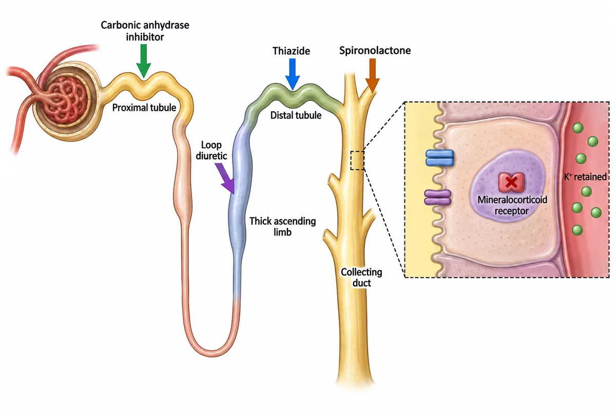
“Spironolactone is a potassium-sparing diuretic” is a fact you can memorise. Understanding where it acts and why potassium is retained gives that fact a structure.
 View larger ↗⤢ Tap to enlarge
The diagram lets you move through three connected ideas: location → receptor → effect. Spironolactone blocks the mineralocorticoid receptor in the distal nephron, including the collecting duct, reducing aldosterone-mediated potassium excretion. The inset makes a cellular mechanism visible within the larger organ map. Mechanism reference: NCBI Bookshelf
This is why a useful zoom is more than a bigger picture. It directs attention to the detail needed to understand the answer while preserving its context. Simply enlarging a cluttered image does not automatically improve memory, and no universal “several times better” memory gain can be promised.
Try it: study the whole nephron, enlarge the inset, then close the image. Name the receptor, locate the site of action and explain the effect on potassium in one sentence. You are now testing whether you can reconstruct the idea. Then try it on more NEET PG pharmacology questions.
2. See relationships instead of memorising isolated names
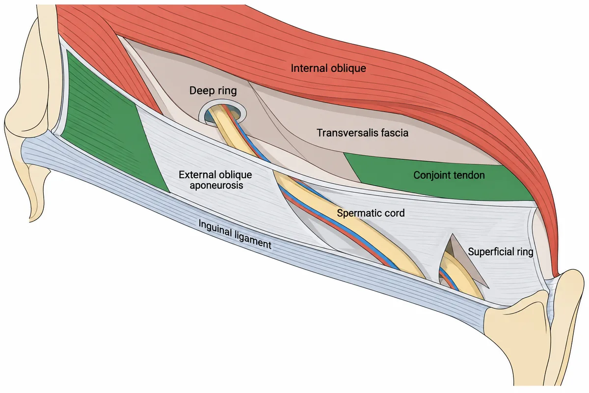
Anatomy becomes difficult when every structure is learned as a separate line of text. The inguinal canal is a good example: anterior wall, posterior wall, roof, floor, deep ring and superficial ring can become a jumble of familiar words.
 View larger ↗⤢ Tap to enlarge
In the illustration, you can follow the spermatic cord through the canal and relate each labelled layer to its neighbours. The posterior wall is transversalis fascia, reinforced medially by the conjoint tendon; the floor is the inguinal ligament. These are the relationships tested in the source MCQ and across the abdomen and pelvis anatomy questions.
A spatial diagram gives you a way to organise those facts. When an option names the wrong wall, you can ask where that structure belongs in the arrangement you have studied.
Try it: cover the labels and identify the posterior wall and floor. Then sketch the canal from memory using only a few lines. The aim is to reproduce the relationships accurately; artistic detail is optional.
3. Compare confusing patterns side by side
Some topics are difficult because the alternatives sound similar. Reading three separate explanations can leave you recognising every term without being able to distinguish them.
 View larger ↗⤢ Tap to enlarge
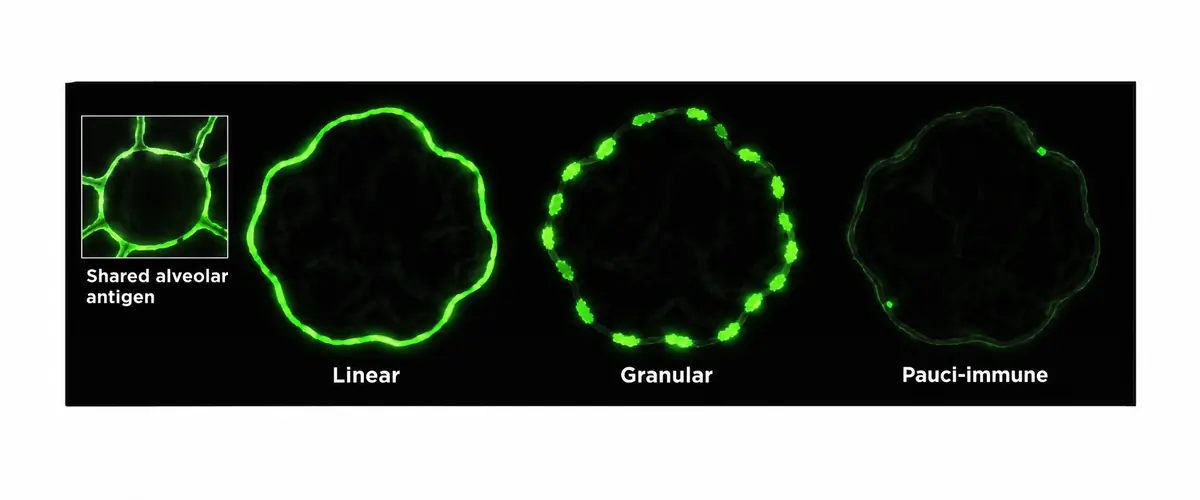
Here, the words linear, granular and pauci-immune become visible differences. Pairing the image with a compact table connects appearance to meaning:
| Pattern | What the pattern suggests | Example association |
|---|---|---|
| Linear | Antibody binding along the basement membrane | Anti-glomerular basement membrane disease |
| Granular | Immune-complex deposition | Lupus nephritis or post-infectious glomerulonephritis |
| Little or no staining | Pauci-immune pattern | ANCA-associated glomerulonephritis |
These are learning associations; a clinical diagnosis also depends on the rest of the findings. The table follows the comparison in Recallab question 50950, one of the nephrology and renal medicine questions.
An image and a table serve complementary purposes. The image shows the pattern. The table aligns the same features across alternatives, making a precise comparison easier. Tables are especially useful when you repeatedly confuse two options: choose a few distinguishing features and compare them in matching rows.
Try it: cover the labels and name each pattern. Then hide the table’s final column and supply an association. Finally, explain why the other patterns would not fit the original MCQ.
4. Connect anatomy to function
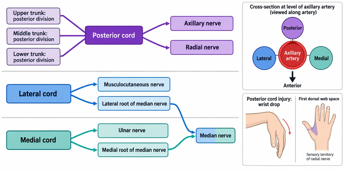
A good explanation can connect facts that otherwise sit in different chapters. A nerve diagram, for example, can bring together anatomical origin, branching and a functional consequence.
 View larger ↗⤢ Tap to enlarge
The posterior cord gives rise to the radial and axillary nerves. The median nerve receives contributions from the lateral and medial cords. Following those branches on the diagram links each name to its origin instead of leaving it as a free-floating fact.
The functional inset provides another question to ask: how could the anatomy help explain a deficit? A finding such as wrist drop needs the full clinical context to localise a lesion; the diagram is a starting point for reasoning, not a shortcut that makes every presentation identical. For the bedside sign that names each nerve, work through the upper limb anatomy questions.
Try it: trace one branch, close the image and describe its origin. Then return to the MCQ and explain which findings support the answer and which help you reject another option.
What does the evidence actually support?
There is a sound educational basis for combining relevant words and pictures. In a study involving medical students, applying multimedia design principles improved short-term retention. That supports careful design—clear organisation, relevant visuals and helpful placement of information. It does not establish that every illustrated MCQ produces lasting recall or a higher NEET PG score. Read the study (Issa et al., Medical Education, 2011)
For remembering over longer periods, retrieval practice matters. A randomised trial in medical education found that repeated testing with feedback produced better long-term retention than repeated, spaced study. This is a reason to use an illustration as something to recall and explain, rather than something to keep rereading. Read the trial (Larsen et al., Medical Education, 2009)
The practical inference is straightforward: use the visual to understand the material, then practise retrieving that understanding over time. These studies support the learning principles; they are not a trial of Recallab or a guarantee of exam-day performance.
How to keep the topic available until your exam
A familiar-looking diagram can give a false sense of mastery. The useful test is what you can explain after you close it.
Build a short routine around illustrated MCQs:
- Answer before opening the explanation. Commit to an option and identify the clue that led you there.
- Study the relevant part of the image. Read its labels, follow its arrows and enlarge small details when needed.
- Close it and retrieve. Name the structures, recreate the comparison or explain the mechanism aloud.
- Check and correct. Reopen the explanation and identify the exact link you missed.
- Return after a gap. A next-day review, another later that week and a later review can be a practical starting pattern. Adjust the gaps to your recall and the time remaining before your exam; this is an example, not a validated universal timetable.
- Change the question. Try an unlabelled view, a differently worded MCQ or a related case. Check that you have learned the concept rather than the location of a label on one page.
As the exam approaches, let your errors guide your visual revision. Revisit the few relationships and patterns you still confuse, and test them again without the image in front of you. In Recallab, Due Today brings questions back for another attempt on a schedule worked out from your own answers.
What makes an MCQ illustration worth your time?
High quality means more than high resolution. The anatomy and mechanism must be correct; labels must remain readable; arrows must point clearly; and the illustration should answer a real learning need. Colour can organise information, but the meaning should also be clear from labels and layout.
A useful image earns its place by helping you locate, compare, explain or recall something. A crowded poster that tries to teach an entire chapter may be less useful than a small, focused diagram. Illustrations also complement practice with authentic clinical images, whose variation and detail a simplified drawing cannot fully reproduce.
For your next revision session, choose one topic you keep forgetting. Attempt a question, study its illustration and close the explanation. If you can redraw the relationship or explain why the competing option is wrong, you have something concrete to practise again tomorrow.
Explore Recallab’s NEET PG question bank and practise with MentorQs, or try a timed NEET PG mock section.
Why candidates use Recallab
- Built towards 22,000 — a batch at a time
- Illustrations drawn for the question
- Every option explained, not just the right one
- AI study tools that are live today
- Start free, today
Or see the plans, or try a timed mock section.
Register to practise NEET PG image MCQsFree account, no card needed. Practise with every option explained and a reference on every question.Drawn from questions #52228, #51804, #50950, #54604 in the Recallab bank, each with its own verified answer and current-edition reference. Last reviewed 19 September 2026.