
Recallab › Blog › Eye and ENT spotters for NEET PG: fundus, lens, eardrum, tuning fork and larynx
Eye and ENT spotters for NEET PG: fundus, lens, eardrum, tuning fork and larynx
Eye and ENT questions give you a picture of one small structure and ask what is wrong with it. The answer is almost always one feature you can learn to look for first.
Register to practise NEET PG eye and ENT questions →Free account, no card needed.
In these two subjects the figure carries the question: a fundus, a lens, an eardrum, a larynx. The pictures below are illustrations drawn for the questions, not photographs of patients. Each is a real Recallab MentorQ, with the feature that decides it. Try each before you open the answer.
Before you name anything: three questions
- Which structure is this? Name the organ and the view, so you know what normal looks like.
- What is the one thing that is not normal? A line, a vessel, a rim, a membrane.
- What does that feature make you do? Treat now, refer, operate, or look for a cause elsewhere.
The eye
Recallab example 1 · retinopathy of prematurity
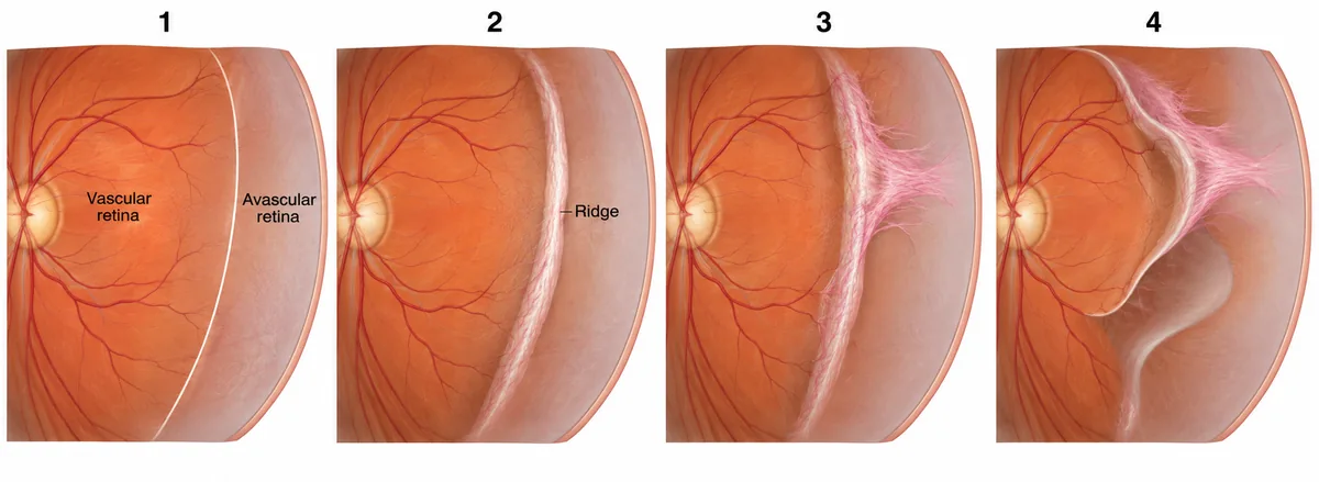
The peripheral retina of a premature infant screened in a special newborn care unit is drawn in the figure in four panels, numbered. Which numbered panel shows stage 3 retinopathy of prematurity?
Look first: follow the border between the vascular and avascular retina from panel 1 on.
 View larger ↗⤢ Tap to enlarge
- APanel 4
- BPanel 1
- CPanel 2
- DPanel 3
Show the answer and explanation
Answer: D. Panel 3
Panel 3. Stage 1 is a flat demarcation line, stage 2 a raised ridge, and stage 3 adds pink fibrovascular tissue growing from the ridge into the vitreous. With plus disease in zone I or II, stage 3 is treated urgently, with laser to the avascular retina or an anti-VEGF injection, to prevent detachment. Screening applies to babies born under 34 weeks or under 2000 grams.
Why the closest rival loses: panel 2 (C) is a ridge with no tissue growing into the vitreous yet. Panel 1 (B) is only the line, and panel 4 (A) is a later stage, with detachment beginning.
Recallab question 55400 · Ophthalmology › Retina · reference: Khurana, Comprehensive Ophthalmology
Recallab example 2 · diabetic fundus
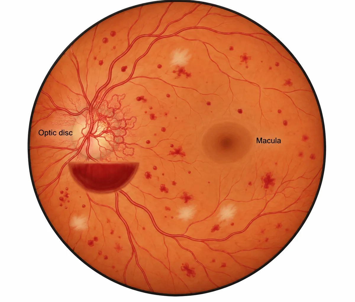
A man who has had diabetes for fifteen years has a visual acuity of 6/9 in the right eye. The fundus of that eye is drawn in the figure. What treatment is due?
Look first: find the optic disc first: what is growing from it, and is there blood?
 View larger ↗⤢ Tap to enlarge
- AReview in six months
- BImmediate vitrectomy
- CFocal macular laser
- DPanretinal photocoagulation
Show the answer and explanation
Answer: D. Panretinal photocoagulation
Panretinal photocoagulation. New vessels on the disc, especially with a preretinal or vitreous haemorrhage, are high-risk proliferative retinopathy: the retina is ischaemic and releasing growth factor. Laser to the peripheral retina reduces that drive and the vessels regress. Good acuity does not delay treatment; the aim is to keep it.
Why the closest rival loses: focal macular laser (C) treats macular oedema, and the macula here is flat. Vitrectomy (B) is for haemorrhage that does not clear, or for traction. Review in six months (A) is for much milder disease.
Recallab question 55991 · Ophthalmology › Retina · reference: Khurana, Comprehensive Ophthalmology
Recallab example 3 · lens opacities
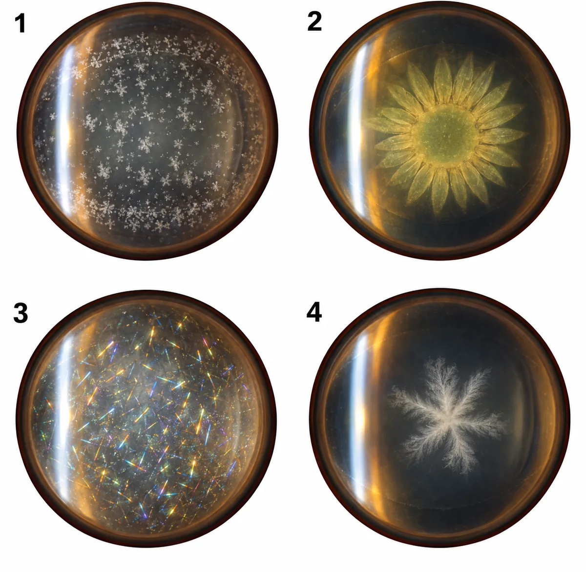
A young man with a tremor and abnormal liver tests has a slit-lamp examination. Lens opacities from four patients are drawn in the figure. Which one fits him?
Look first: a tremor with liver disease in a young man: name the condition before the lens.
 View larger ↗⤢ Tap to enlarge
- AOpacity 3, a Christmas tree cataract
- BOpacity 1, a snowflake cataract
- COpacity 4, a rosette cataract
- DOpacity 2, a sunflower cataract
Show the answer and explanation
Answer: D. Opacity 2, a sunflower cataract
Opacity 2, a sunflower cataract. A tremor and liver disease in a young man mean Wilson disease. Copper deposits under the anterior lens capsule in a petal pattern, the sunflower cataract, and in Descemet's membrane as a Kayser-Fleischer ring at the corneal edge. It rarely affects vision, and both fade with chelation.
Why the closest rival loses: a Christmas tree cataract (A), with multicoloured crystals, belongs to myotonic dystrophy. The snowflake cataract (B) is the diabetic one, and the rosette (C) the traumatic one.
Recallab question 55587 · Ophthalmology › Lens · reference: Khurana, Comprehensive Ophthalmology
Ear, nose and throat
Recallab example 4 · the eardrum
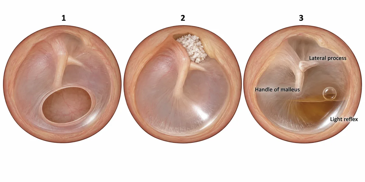
Three tympanic membranes are drawn in the figure. Membrane 2 carries the dangerous type of disease. What is it about the defect that makes it so?
Look first: look at the edge of each defect: is there a rim of membrane all the way round?
 View larger ↗⤢ Tap to enlarge
- AIt lies above the lateral process
- BIt lies behind a fluid level
- CIt is large and central
- DIt reaches the bony margin
Show the answer and explanation
Answer: D. It reaches the bony margin
It reaches the bony margin. A defect with no rim of membrane between it and the bone cannot contain the debris: skin grows in from the edge, sheds keratin into a closed space, and builds a cholesteatoma. That keratin erodes bone and can reach the facial nerve, the labyrinth and the dura, which is why this disease is called unsafe and is treated by surgery.
Why the closest rival loses: a large central defect (C), as in membrane 1, keeps its rim, so the disease stays in the mucosa. A fluid level (B) is an effusion behind an intact drum, as in membrane 3. Position and size (A) are not what makes it dangerous; the missing rim is.
Recallab question 55044 · E.N.T › Ear · reference: Dhingra, Diseases of Ear, Nose and Throat
Recallab example 5 · tuning fork tests
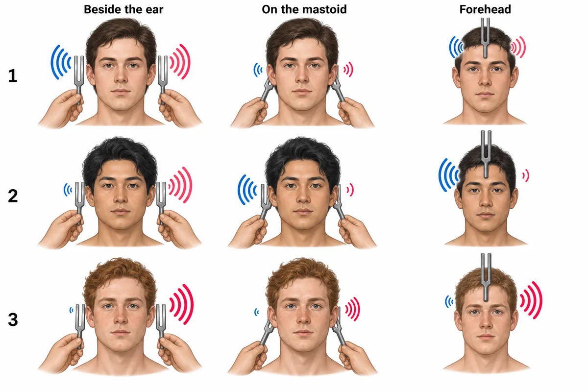
Three patients' tuning fork results are drawn in the figure as pictures rather than words. One patient has a conductive loss in the right ear. What does the Weber test show in that patient?
Look first: remember the picture is a face: the patient's right ear is on your left.
 View larger ↗⤢ Tap to enlarge
- ALouder in the better ear
- BHeard centrally
- CLouder on bone than air
- DLouder in the affected ear
Show the answer and explanation
Answer: D. Louder in the affected ear
Louder in the affected ear. In a conductive loss, bone conduction is better than air on the affected side, and the blocked ear is shielded from background noise, so the fork on the forehead is heard louder in that ear. That is patient 2. The left ear behaves normally, with air better than bone.
Why the closest rival loses: a Weber heard louder in the better ear (A) is the sensorineural pattern, patient 3. A central Weber (B) is patient 1, with symmetrical hearing. Bone louder than air (C) describes the Rinne test, not the Weber.
Recallab question 55141 · E.N.T › Ear · reference: Dhingra, Diseases of Ear, Nose and Throat
Recallab example 6 · the infant larynx
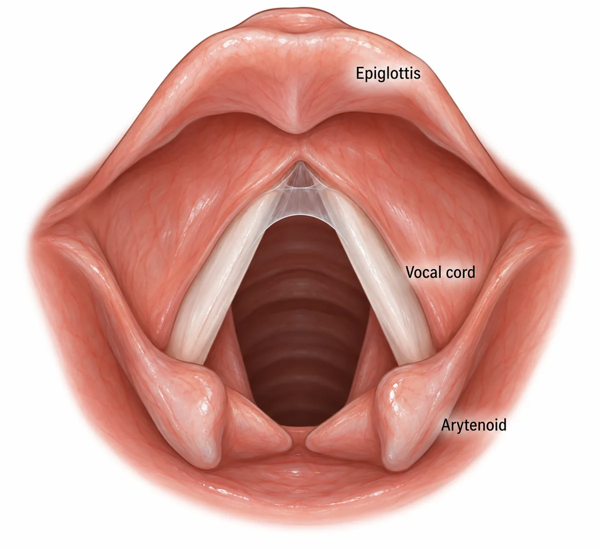
The larynx of an infant with a weak, hoarse cry and noisy breathing since birth is drawn in the figure as seen at laryngoscopy. What is the lesion?
Look first: start at the front of the cords, where they meet, and look for anything between them.
 View larger ↗⤢ Tap to enlarge
- ACongenital anterior glottic web
- BLaryngomalacia
- CBilateral vocal cord palsy
- DSubglottic haemangioma
Show the answer and explanation
Answer: A. Congenital anterior glottic web
Congenital anterior glottic web. The larynx is closed by epithelium in early fetal life and later reopens. If reopening fails at the front, a web remains between the cords. It limits cord vibration, giving a weak or hoarse cry from birth, and a large web narrows the airway, adding stridor. Anterior webs are linked to the 22q11.2 deletion syndrome, so cardiac and genetic review follow.
Why the closest rival loses: laryngomalacia (B) shows a curled epiglottis and prolapsing arytenoids, and the cry is normal. In bilateral cord palsy (C) the cords lie near the midline, not open with a membrane between them.
Recallab question 55643 · E.N.T › Larynx · reference: Dhingra, Diseases of Ear, Nose and Throat
The one-minute routine
| If you see | Look first | It usually settles |
|---|---|---|
| A premature infant's retina | A line, a ridge, or tissue growing inwards? | The stage of ROP, and whether to treat now |
| A diabetic fundus | New vessels on the disc, and any blood? | Proliferative disease: panretinal laser |
| A slit-lamp lens opacity | What shape is it? | The cause: copper, diabetes, trauma, myotonic dystrophy |
| An eardrum | Is there a rim of membrane all the way round? | Safe or unsafe disease; surgery for unsafe |
| Tuning fork results | Bone better than air on which side? | Conductive or sensorineural loss |
| An infant's larynx | Is anything between the cords? | A web, and the 22q11 deletion to look for |
For other kinds of figure, see ten NEET PG image questions and forensic medicine spotters. For sections through the limbs, trunk, neck and brain, see six anatomy cross-sections and surgical instruments and procedures. To practise the topics that matter most one at a time, see high impact topics for NEET PG.
For skin pictures and nail signs, see six NEET PG dermatology questions.
Why candidates use Recallab
- Built towards 22,000 — a batch at a time
- Illustrations drawn for the question
- Every option explained, not just the right one
- AI study tools that are live today
- Start free, today
Or see the plans, or try a timed mock section.
Register to practise NEET PG eye and ENT questionsFree account, no card needed. Practise with every option explained and a reference on every question.Drawn from questions #55400, #55991, #55587, #55044, #55141, #55643 in the Recallab bank, each with its own verified answer and current-edition reference. Last reviewed 4 October 2026.