
Recallab › Blog › Surgical instruments and procedures for NEET PG: six tools you can name from their shape
Surgical instruments and procedures for NEET PG: six tools you can name from their shape
An instrument question is a question about a job. Once you ask what the tool has to do, its shape usually tells you its name.
Register to practise NEET PG surgery questions →Free account, no card needed.
Instrument and procedure questions show a tray, a row of retractors or a tube and ask which one fits the job. The pictures below are illustrations drawn for the questions, not photographs. Each is a real Recallab MentorQ, with the feature that decides it. Try each before you open the answer.
Before you name a tool: three questions
- What is the job? Hold open, hold back, measure, drain, remove a clot, keep an airway open. One verb narrows the field.
- Where does it go, and for how long? A tool for a duct, a vessel, the airway or a body cavity is built differently from one for skin.
- What would go wrong if it were the wrong one? A blade that cuts, a tube that blocks, a tip that perforates. The wrong options usually fail in just that way.
In theatre
Recallab example 1 · the evacuation tray
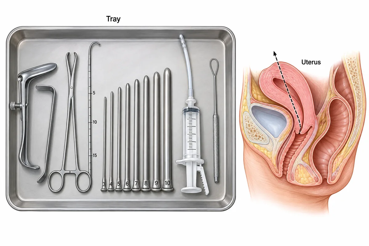
An instrument tray is drawn in the figure laid out for surgical evacuation of the uterus. Which instrument is passed first into the uterine cavity, to measure its length and direction?
Look first: read the tray as an order of use, then ask which instrument measures before anything is dilated.
 View larger ↗⤢ Tap to enlarge
- AThe speculum
- BThe looped instrument
- CThe rod marked in centimetres
- DThe toothed forceps
Show the answer and explanation
Answer: C. The rod marked in centimetres
The rod marked in centimetres: the uterine sound. Before the cavity is dilated, the operator needs its depth and its direction. The sound gives both: its centimetre marks read the length and its curve shows the direction. That length then limits how far the dilators and the cannula are passed. It goes in blind, so a misjudged angle can take it through the uterine wall, as the dotted line shows.
Why the closest rival loses: the looped instrument (B) is the curette, used last, once the depth is known. The toothed forceps (D) is the tenaculum: it grasps and steadies the cervix and never enters the cavity. The speculum (A) shows the cervix and stays in the vagina. Pulling the cervix down with the tenaculum straightens the angle between cervix and body, which is what lets the sound follow the canal rather than the fundus.
Recallab question 55195 · Obstetrics › Early Pregnancy Complications · reference: DC Dutta's Textbook of Gynecology
Recallab example 2 · the liver retractor
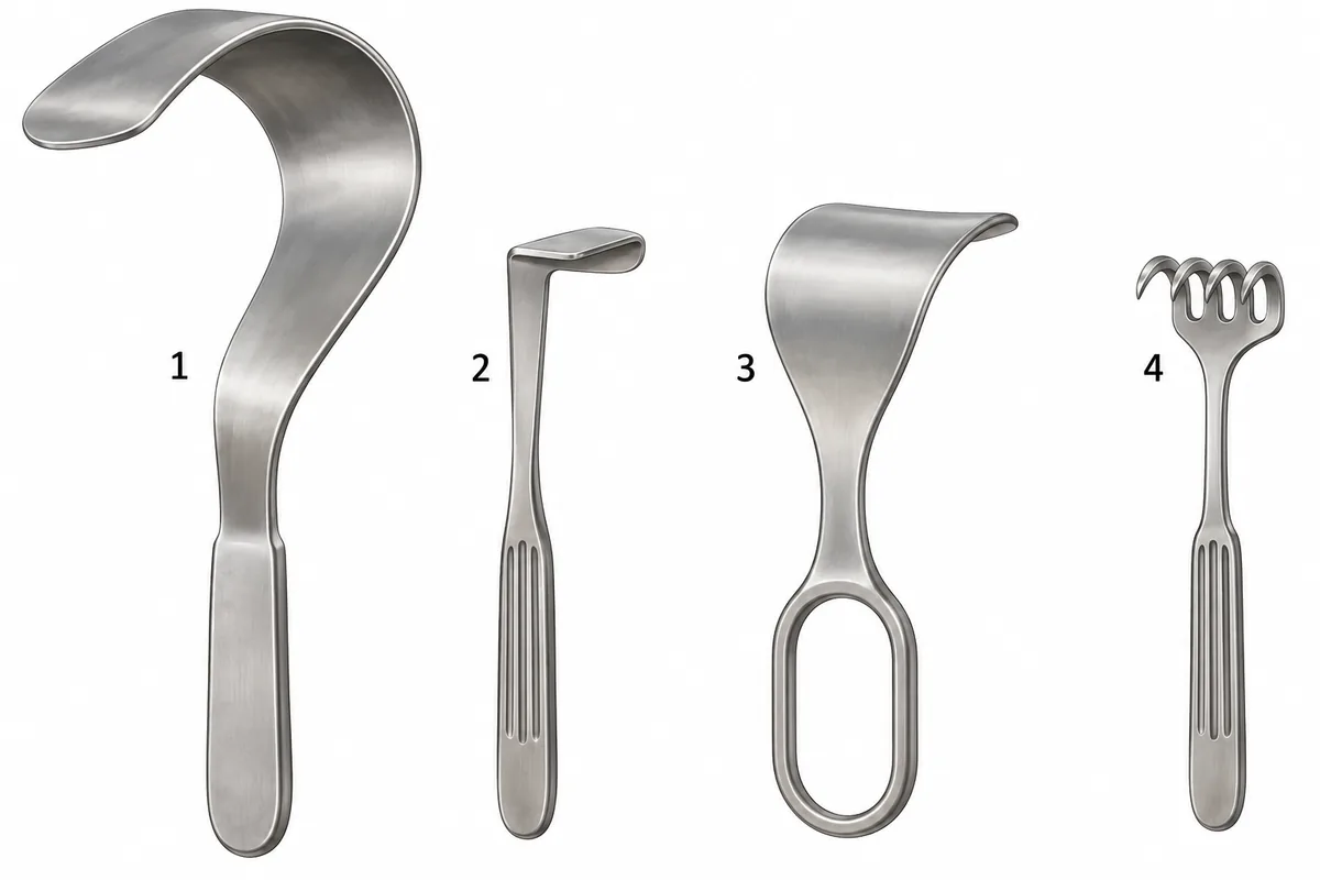
During an upper abdominal operation the assistant must hold the edge of the liver up and out of the field for a long time. Four hand-held retractors are drawn in the figure. Which one is designed for that?
Look first: compare how long and how deeply curved each blade is against how far down the liver lies.
 View larger ↗⤢ Tap to enlarge
- ARetractor 3
- BRetractor 2
- CRetractor 4
- DRetractor 1
Show the answer and explanation
Answer: D. Retractor 1
Retractor 1, the Deaver. Its long, broad blade curves deeply, like a question mark. The curve hooks under a solid organ and holds it up, and the smooth blade spreads the pressure so the liver is not cut. Its length reaches deep into the upper abdomen, under the liver or the costal margin.
Why the closest rival loses: retractor 3 (A) is broad, but only shallowly curved, a retractor for the abdominal wall rather than a deep hook for an organ. Retractor 2 (B) is small and right-angled, for skin and superficial layers. Retractor 4 (C) is a sharp rake that grips skin edges and would tear liver tissue.
Recallab question 56062 · Surgery › General Surgery Principles · reference: Bailey & Love's Short Practice of Surgery
Recallab example 3 · the bile duct drain
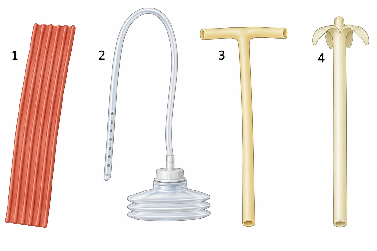
After the common bile duct is opened and cleared of stones, a drain is left inside the duct itself. Four drains are drawn in the figure. Which one is used?
Look first: ask which drain sits inside a duct, and which only lies beside an organ or in a wound.
 View larger ↗⤢ Tap to enlarge
- ADrain 1
- BDrain 2
- CDrain 3
- DDrain 4
Show the answer and explanation
Answer: C. Drain 3
Drain 3, the T-tube. Its short limbs lie inside the duct and its long limb leaves through the skin. It decompresses the duct while the opening heals and the swelling at the papilla settles. Contrast injected through it shows whether a stone has been left behind, and a normal cholangiogram with free flow into the duodenum is the signal to remove it, at about ten to fourteen days, by which time a fibrous track has formed.
Why the closest rival loses: drain 2 (B) is a closed suction drain, which lies beside an organ in the peritoneal space, never inside a duct. Drain 1 (A) is a corrugated sheet drain for a wound or the peritoneal space. Drain 4 (D) is a mushroom-tipped catheter of the Malecot kind, for hollow organs such as the bladder.
Recallab question 56060 · Surgery › The Gall Bladder & Bile Ducts · reference: Bailey & Love's Short Practice of Surgery
Vessels, the airway and the blade
Recallab example 4 · the embolectomy catheter
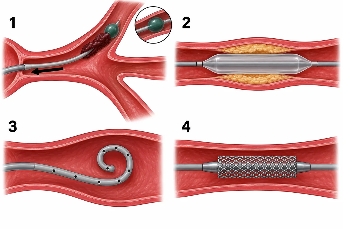
A woman in atrial fibrillation has had a cold, painful, pulseless leg for three hours. Four catheters are drawn in the figure. Which catheter is used to remove the cause?
Look first: name what is blocking the artery first, then ask which catheter can reach beyond it.
 View larger ↗⤢ Tap to enlarge
- ACatheter 4
- BCatheter 2
- CCatheter 3
- DCatheter 1
Show the answer and explanation
Answer: D. Catheter 1
Catheter 1, the Fogarty balloon catheter. Atrial fibrillation with a sudden cold, painful, pulseless leg means an embolus. The catheter is passed through an arteriotomy beyond the clot, its balloon is inflated and it is withdrawn, dragging the clot out. Heparin is given at once and the source is anticoagulated.
Why the closest rival loses: catheter 2 (B) is an angioplasty balloon, which dilates a chronic stenosis and does not remove an embolus. Catheter 4 (A) carries a stent, which holds a narrowing open. Catheter 3 (C) is a pigtail, which delivers contrast for angiography and does not clear clot.
Recallab question 55478 · Surgery › Arterial Disorders · reference: Bailey & Love's Short Practice of Surgery
Recallab example 5 · the tracheostomy tube
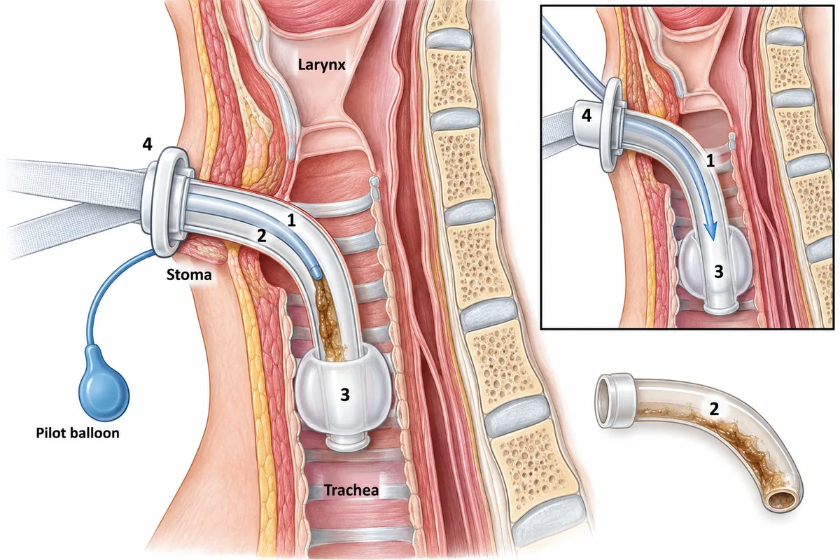
A tracheostomy tube is drawn in the figure in position with four parts numbered. A patient with this tube becomes breathless and the suction catheter will not pass. Which numbered part is the likeliest cause?
Look first: find the part that is made to come out, and ask what collects inside it.
 View larger ↗⤢ Tap to enlarge
- APart 1
- BPart 4
- CPart 2
- DPart 3
Show the answer and explanation
Answer: C. Part 2
Part 2, the inner tube. Dried secretions collect in the inner tube, which is why the tube has one. A catheter that will not pass means the lumen is blocked, and taking the inner tube out is quick, reversible and restores the full lumen of the outer tube. The side panel shows the catheter passing freely once it is out.
Why the closest rival loses: part 1 (A) is the outer tube, and removing it is the next step, not the first, because it risks losing an immature track. Part 3 (D) is the cuff: deflating it does not clear a blocked lumen. Part 4 (B) is the tapes, which when loose or tight cause displacement, not obstruction. If the catheter still will not pass after the inner tube is out, the tube has probably left the trachea, and the emergency steps follow.
Recallab question 55142 · E.N.T › Larynx and Neck · reference: Diseases of Ear, Nose and Throat
Recallab example 6 · the scalpel blade
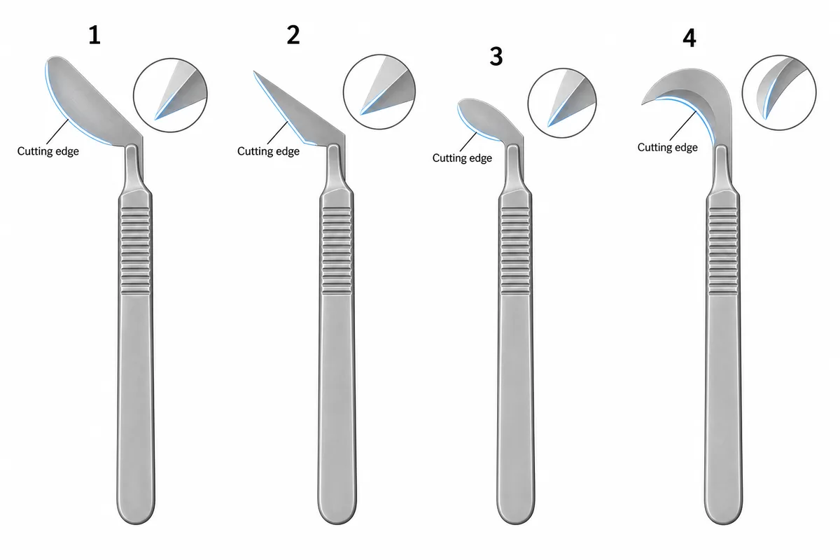
Four detachable blades are drawn in the figure in profile at one scale on the same handle. Which blade is chosen to enter a deep structure point first?
Look first: ask whether each blade is made to go in point first, or to be drawn along a surface.
 View larger ↗⤢ Tap to enlarge
- ABlade 4
- BBlade 1
- CBlade 2
- DBlade 3
Show the answer and explanation
Answer: C. Blade 2
Blade 2. A stab is made point first with a straight-edged pointed blade, and the cut is then extended by drawing the edge outwards. Blade 2's straight edge runs to a sharp forward point, with the edge underneath, so it enters by penetration, to a controlled depth. It is the blade reached for to open an abscess, a vessel or a sheath.
Why the closest rival loses: blade 1 (B), with the long curved belly, is the general skin blade, drawn along the surface rather than pushed in. Blade 3 (D) is a small curved blade for fine, close work. Blade 4 (A) is the hooked crescent, used to cut something already caught in the hook, such as a stitch.
Recallab question 56562 · Surgery › General Surgery Principles · reference: Bailey & Love's Short Practice of Surgery
The one-minute routine
| If you see | Ask first | It usually settles |
|---|---|---|
| A tray laid out for a procedure | Which instrument goes in first, and why | The order of use: measure, dilate, then clear |
| Four retractors | How deep, and what is it holding back | Organ or wall |
| Four drains | Does it sit in a duct, a wound or a space | Which drain, and when it comes out |
| Four catheters in an artery | What is blocking it, and what must pass beyond | Remove, dilate, hold open or image |
| A tube in the neck | Which part is made to come out | Obstruction or displacement |
| Four scalpel blades | Is the edge straight or curved, and where does it start | Stab (point first) or draw (belly) |
For other kinds of figure, see ten NEET PG image questions and six eye and ENT spotters. For the decision that follows the picture, see next-best-step management questions.
For the films that follow an injury, see six fracture and radiograph questions.
Why candidates use Recallab
- Built towards 22,000 — a batch at a time
- Illustrations drawn for the question
- Every option explained, not just the right one
- AI study tools that are live today
- Start free, today
Or see the plans, or try a timed mock section.
Register to practise NEET PG surgery questionsFree account, no card needed. Practise with every option explained and a reference on every question.Drawn from questions #55195, #56062, #56060, #55478, #55142, #56562 in the Recallab bank, each with its own verified answer and current-edition reference. Last reviewed 5 October 2026.