
Recallab › Blog › Fractures and radiographs for NEET PG: six questions and the line to find first
Fractures and radiographs for NEET PG: six questions and the line to find first
A fracture question is rarely about the crack. It is about what the crack does next: to the joint, to the blood supply, to the growth plate.
Register to practise NEET PG orthopaedics questions →Free account, no card needed.
Orthopaedic questions show a drawn film and ask you to name the fracture, the classification grade or the next decision. The pictures below are illustrations drawn for the questions, not patient films. Each is a real Recallab MentorQ, and each answer turns on one feature of the drawing. Try each before you open the answer.
Before you name a fracture: three questions
- Does the line enter the joint? An intra-articular fracture is judged by how well the surface can be put back together.
- What happened to the blood supply? The head of the femur, the head of the humerus and the waist of the dens all depend on a thin supply that a break can cut.
- Is the patient still growing? A growth plate changes both the pattern of the fracture and what must be fixed.
The arm
Recallab example 1 · the distal radius
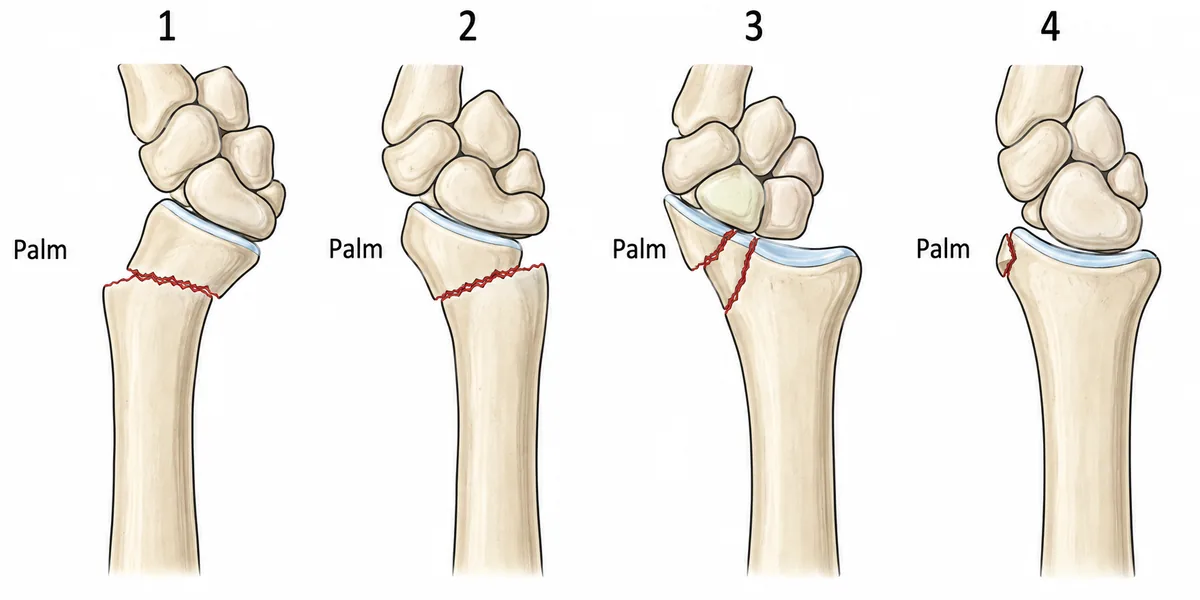
Four lateral drawings of the distal radius after falls are shown in the figure. Which is a volar Barton fracture?
Look first: ask first whether the line runs into the joint, then which way the carpus has gone.
 View larger ↗⤢ Tap to enlarge
- ADrawing 4
- BDrawing 1
- CDrawing 2
- DDrawing 3
Show the answer and explanation
Answer: D. Drawing 3
Drawing 3. A volar Barton fracture splits off the palmar rim of the radius and runs into the joint, and the carpus slips palmarwards together with that fragment. It is a fracture-subluxation of the radiocarpal joint. The rim fragment is small, but the whole carpus rests on it, so plaster cannot hold it: the carpus pushes the fragment away again. It is fixed open with a palmar buttress plate, which also restores the joint surface and limits later arthritis.
Why the closest rival loses: drawing 2 (C) is an extra-articular Smith fracture: the fragment tilts towards the palm, but the joint surface is intact. Drawing 1 (B) is a Colles fracture, tilted towards the back of the hand, and it also spares the joint. Drawing 4 (A) is a fracture of the tip of the radial styloid, the chauffeur fracture, which does not carry the carpus away.
Recallab question 55246 · Orthopaedic › Injuries Around The Fore Arm & Wrist · reference: Essential Orthopaedics
Recallab example 2 · the proximal humerus
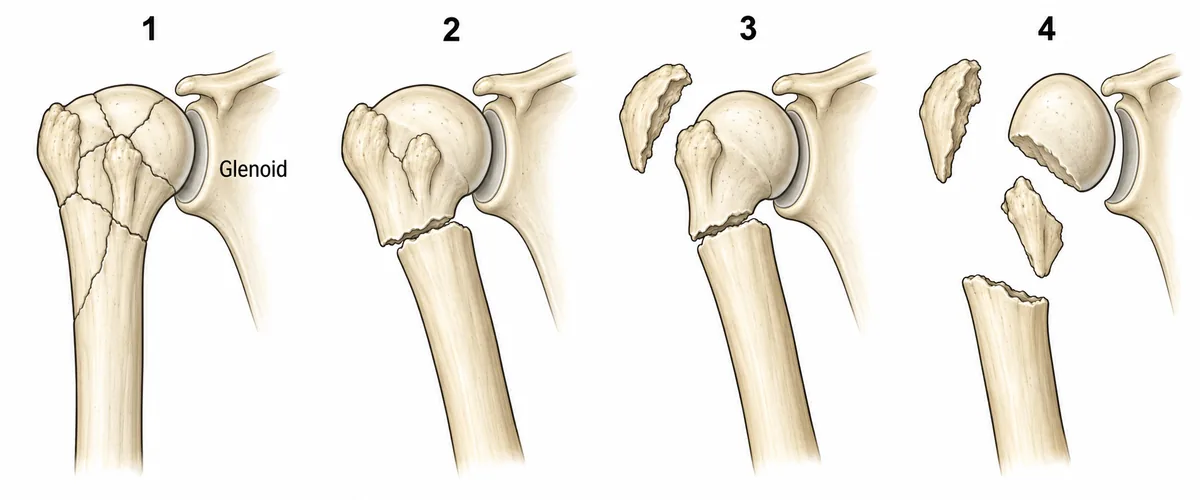
A woman of 72 fell on her outstretched hand. Four patterns of proximal humerus fracture are drawn in the figure, and hers is one of them. Which pattern most threatens the blood supply of the humeral head?
Look first: count the displaced parts, not the lines, then ask what the head is still joined to.
 View larger ↗⤢ Tap to enlarge
- APattern 3
- BPattern 4
- CPattern 1
- DPattern 2
Show the answer and explanation
Answer: B. Pattern 4
Pattern 4. Neer's classification counts displaced parts, not fracture lines: the head, the two tuberosities and the shaft. A part counts only when it is displaced more than 1 cm or angulated more than 45 degrees. In pattern 4 the head is left attached to nothing, so the vessels it draws from the circumflex humeral arteries are torn, and avascular necrosis is likeliest. In an older patient this often leads to arthroplasty rather than fixation.
Why the closest rival loses: pattern 3 (A) is a three-part fracture: the head is still joined to the lesser tuberosity, so some of its supply survives. Pattern 1 (C) has many lines but nothing displaced, so it counts as one part. Pattern 2 (D) is a two-part neck fracture, and the head keeps both tuberosities and their soft tissue.
Recallab question 56166 · Orthopaedic › Injuries Around Shoulder · reference: Apley & Solomon's System of Orthopaedics and Trauma
The hip and the neck
Recallab example 3 · the femoral neck
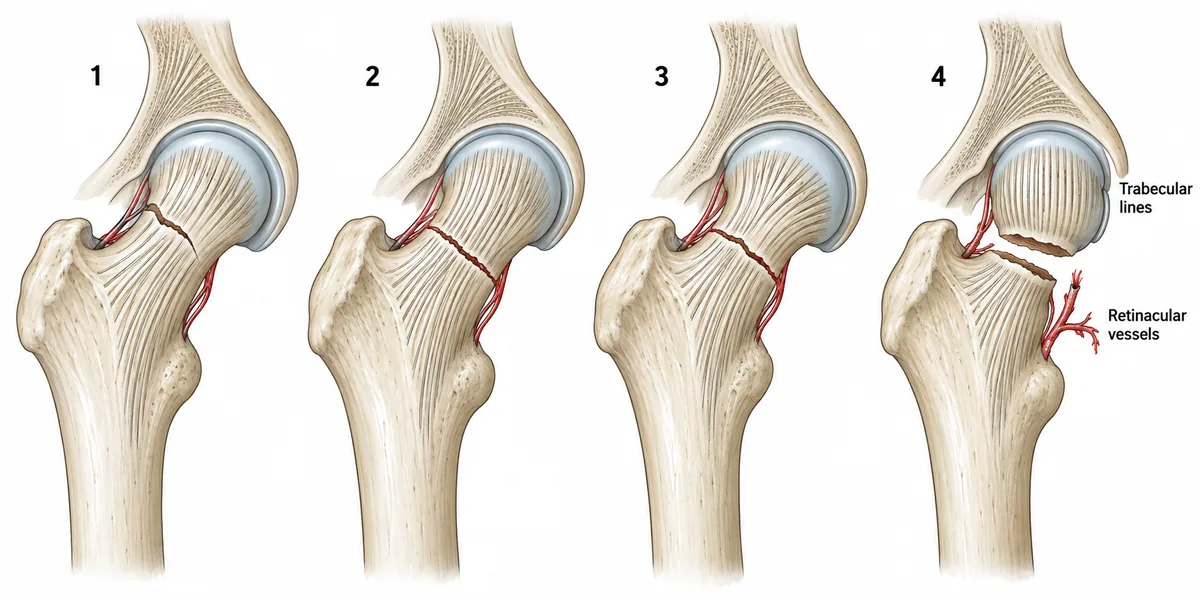
Four femoral neck fractures are drawn in the figure with the trabecular lines of the head and neck marked in each. Which drawing shows a Garden grade four?
Look first: compare how the fine lines of the head and neck match up, and read those, not the gap.
 View larger ↗⤢ Tap to enlarge
- ADrawing 1
- BDrawing 2
- CDrawing 4
- DDrawing 3
Show the answer and explanation
Answer: C. Drawing 4
Drawing 4. Garden grade four is a complete fracture with full displacement. Full displacement tears the retinacular vessels that run up the neck, on which most of the head's blood supply depends, so the head is left relying on a small foveal branch. That is why a displaced fracture in an older patient is replaced, not fixed. The trap: with the head freed completely, it lies parallel to the acetabulum again, so grade four can look less deformed than grade three.
Why the closest rival loses: drawing 3 (D) is grade three, partly displaced, the head rotated but the retinaculum intact. Drawing 2 (B) is grade two: complete but undisplaced, with the trabeculae lining up throughout. Drawing 1 (A) is grade one, incomplete and impacted in valgus, the most stable.
Recallab question 55045 · Orthopaedic › Spine, Pelvis & Lower Limb Trauma · reference: Apley & Solomon's System of Orthopaedics and Trauma
Recallab example 4 · the axis
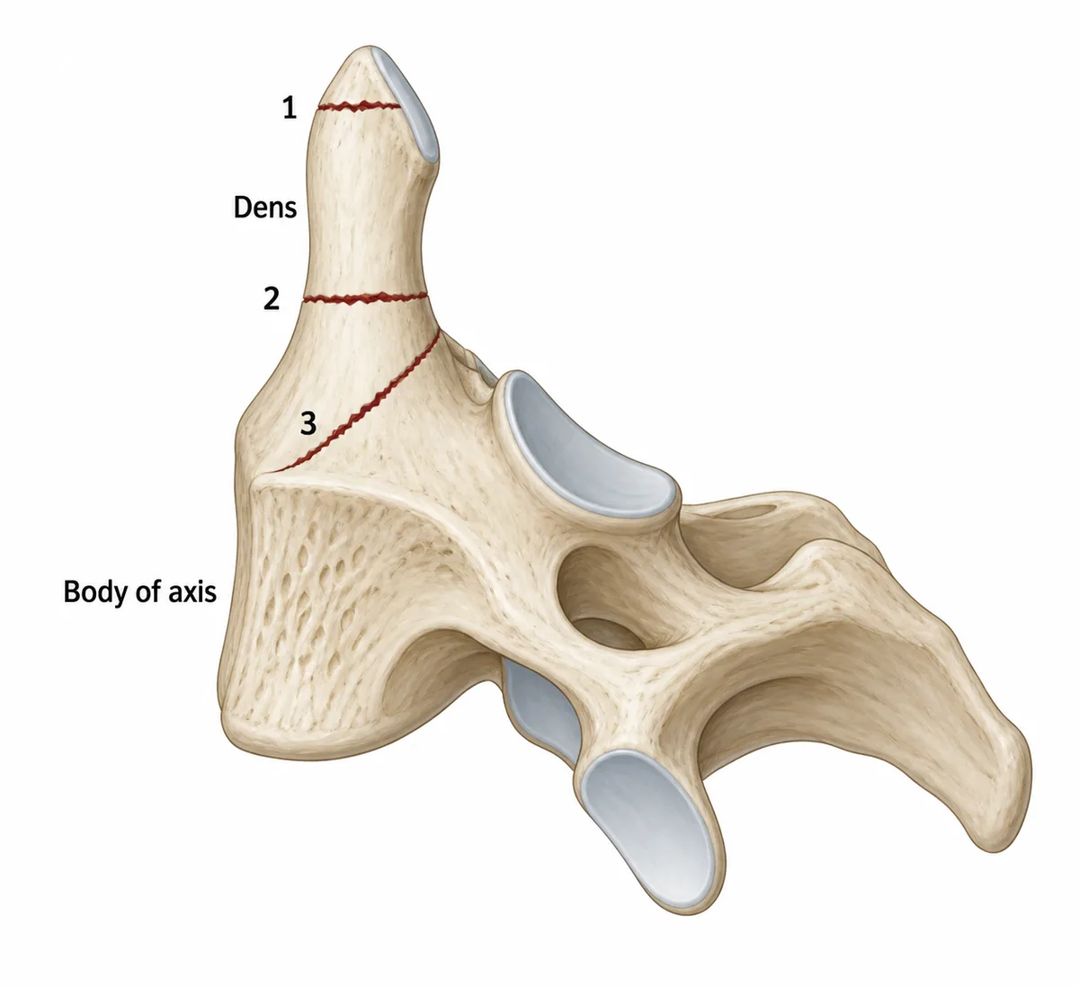
Three fracture lines of the axis are drawn in the figure, numbered. Which numbered fracture has the highest rate of non-union?
Look first: name the part of the bone each line crosses, then ask which part has the poorest blood supply.
 View larger ↗⤢ Tap to enlarge
- AFracture 3, into the body
- BHangman fracture of the pedicles
- CFracture 2, the dens waist
- DFracture 1, the dens tip
Show the answer and explanation
Answer: C. Fracture 2, the dens waist
Fracture 2, across the waist of the dens. That segment has a poorer blood supply than the base or the tip, and the fracture surfaces have a small area of bone-to-bone contact. Together those give it the highest non-union rate of the three. It often needs anterior screw fixation or a posterior fusion, especially in older patients.
Why the closest rival loses: fracture 3 (A) runs into the vascular cancellous body of the axis, which unites reliably in a collar or halo. Fracture 1 (D), through the tip, is a stable avulsion injury that unites well with external support. A hangman fracture (B) goes through the pedicles of C2, which is not one of the drawn lines, and it unites well in a halo.
Recallab question 55372 · Orthopaedic › Spinal Injuries · reference: Essential Orthopaedics
The hand and the growing bone
Recallab example 5 · the thumb base
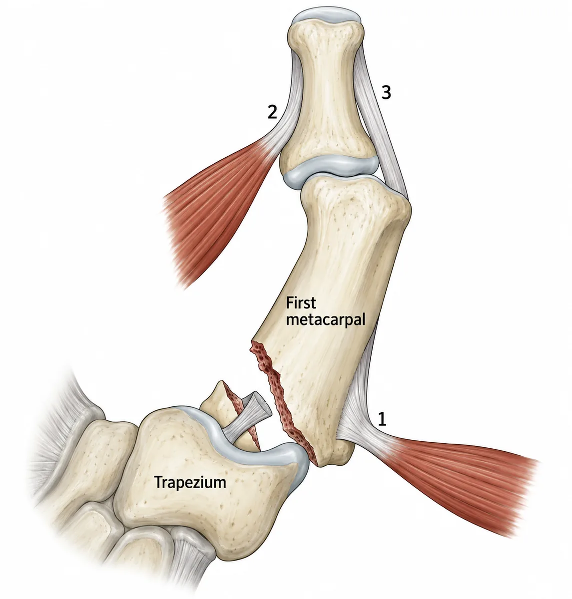
A boxer has a painful swollen thumb base after a punch. The fracture and three tendons attaching near it are drawn in the figure. Which structure displaces the main fragment?
Look first: find the small fragment still tied to the trapezium, then ask which tendon pulls the shaft away from it.
 View larger ↗⤢ Tap to enlarge
- ATendon 2
- BTendon 1
- CTendon 3
- DThe ligament to the trapezium
Show the answer and explanation
Answer: B. Tendon 1
Tendon 1, abductor pollicis longus. The oblique fracture runs into the joint and splits off a small fragment that stays tethered to the trapezium by the volar oblique ligament: a Bennett fracture. Tendon 1 inserts on the base of the shaft fragment itself, and its pull draws the shaft upwards and outwards off the trapezium, with adductor pollicis adding to the displacement. The joint surface is split, so fixation is usually needed.
Why the closest rival loses: tendon 2 (A) is adductor pollicis, which adds adduction, but the proximal pull on the shaft is from abductor pollicis longus. Tendon 3 (C), extensor pollicis brevis, inserts on the proximal phalanx and acts across the next joint. The ligament (D) holds the small fragment against the trapezium and displaces nothing.
Recallab question 55767 · Orthopaedic › Hand Injuries · reference: Apley & Solomon's System of Orthopaedics and Trauma
Recallab example 6 · the teenager's ankle
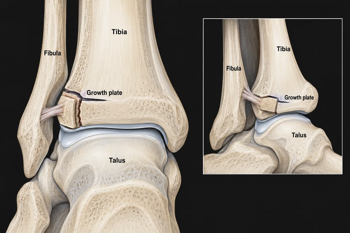
A 14-year-old girl twisted her foot outwards while skating. Her ankle radiographs are drawn in the figure. What is the injury?
Look first: find the growth plate, see which part of it has closed, then follow the fracture line.
 View larger ↗⤢ Tap to enlarge
- ASalter-Harris type II fracture
- BJuvenile Tillaux fracture
- CTriplane fracture
- DSalter-Harris type I separation
Show the answer and explanation
Answer: B. Juvenile Tillaux fracture
A juvenile Tillaux fracture. The distal tibial growth plate closes over about eighteen months in the early teens, starting centrally and medially and finishing at the front outer corner. In that window only the anterolateral part of the epiphysis is still free. An outward twist pulls on the anterior tibiofibular ligament attached there, and the ligament pulls that corner off, the fracture line running down through the epiphysis into the joint. It is a Salter-Harris type III injury, fixed if the step in the joint exceeds two millimetres.
Why the closest rival loses: a type II fracture (A) runs along the plate and out through the metaphysis above it, but this one runs down into the joint. A triplane fracture (C) adds a fragment of bone above the plate on the lateral view, and the inset shows none. A type I separation (D) cannot be right: the medial part of the plate has already fused and the fracture enters the joint.
Recallab question 55965 · Orthopaedic › Injuries To The Leg Ankle & Foot · reference: Apley & Solomon's System of Orthopaedics and Trauma
The one-minute routine
| If you see | Ask first | It usually settles |
|---|---|---|
| Four drawings of the wrist | Does the line enter the joint, and which way is the carpus | Which named fracture, and whether plaster will hold it |
| Four patterns at the shoulder | How many parts are displaced | Whether the head keeps its supply |
| Four fractures of the hip | Are the head and neck lines still lined up | Displaced or undisplaced, and so fix or replace |
| A fracture through the axis | Which level of the peg | How well it will unite |
| A fracture at the thumb base | Which part is still tied to the trapezium | Which tendon pulls the shaft away |
| A teenager's ankle | Which part of the growth plate has closed | The pattern, and whether the joint surface has a step |
For other kinds of figure, see ten NEET PG image questions and six eye and ENT spotters. For the decision that follows the picture, see next-best-step management questions.
For skin pictures and nail signs, see six NEET PG dermatology questions.
Why candidates use Recallab
- Built towards 22,000 — a batch at a time
- Illustrations drawn for the question
- Every option explained, not just the right one
- AI study tools that are live today
- Start free, today
Or see the plans, or try a timed mock section.
Register to practise NEET PG orthopaedics questionsFree account, no card needed. Practise with every option explained and a reference on every question.Drawn from questions #55246, #56166, #55045, #55372, #55767, #55965 in the Recallab bank, each with its own verified answer and current-edition reference. Last reviewed 6 October 2026.