
Recallab › Blog › Anatomy cross-sections for NEET PG: six sections and the one thing to find first
Anatomy cross-sections for NEET PG: six sections and the one thing to find first
A cross-section looks crowded until you know where to start. In every one, the answer is a relation: what lies beside a bone, inside a fascial layer, or between two landmarks.
Register to practise NEET PG anatomy questions →Free account, no card needed.
Anatomy sections turn up as a numbered picture and a short question: which nerve, which muscle, which space. The pictures are illustrations drawn for the questions. Below are six real Recallab MentorQs, one for each region from the limbs to the brain. Try each before you open the answer.
Before you read any section: three questions
- What level is this? Name it from the bone: a humerus, the carpal row, a lumbar vertebra, a cervical vertebra, the thalamus.
- Where are the fixed landmarks? A septum, a fascial layer, a big vessel. They stay put, so everything else is described from them.
- Which side is which? Most sections are drawn as seen from below, so the patient's right is on your left. Check the labels before you trust your instinct.
The limbs
Recallab example 1 · the arm
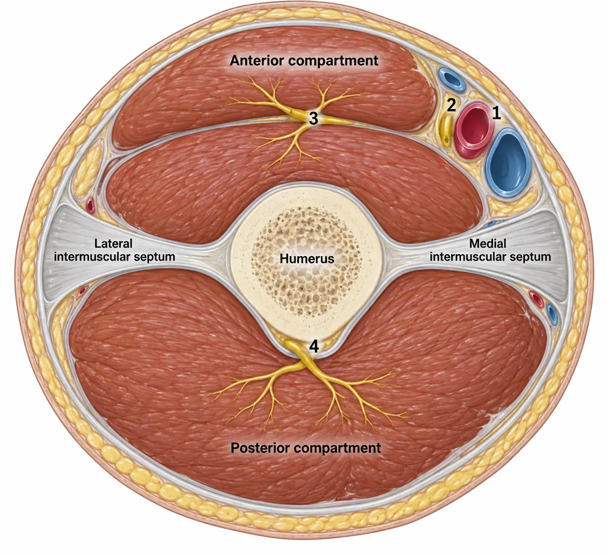
A transverse section through the middle of the arm is drawn in the figure with four structures numbered. Which nerve is structure 3?
Look first: find the two compartments first, then ask which nerve runs between the muscles and branches into them.
 View larger ↗⤢ Tap to enlarge
- AMedian nerve
- BUlnar nerve
- CRadial nerve
- DMusculocutaneous nerve
Show the answer and explanation
Answer: D. Musculocutaneous nerve
The musculocutaneous nerve. The arm has two compartments, separated by the two septa. Structure 3 lies between the two flexor bellies and branches into both, so it supplies the anterior compartment: the musculocutaneous nerve, which flexes the elbow. Structure 2 passes through the arm giving nothing: the median nerve. Structure 4, in the groove behind the bone, is the radial nerve.
Why the closest rival loses: the median nerve (A) is structure 2 and gives no branch in the arm. The ulnar nerve (B) pierces the medial septum into the back compartment and also gives no branch here. The radial nerve (C) is structure 4, in the spiral groove.
Recallab question 55004 · Anatomy › Upper Limb · reference: Gray's Anatomy for Students
Recallab example 2 · the wrist
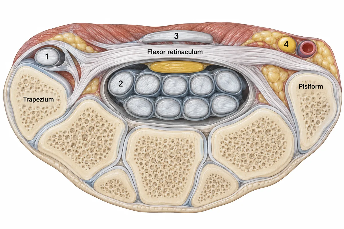
A transverse section of the right wrist at the level of the distal carpal row is drawn in the figure with four structures numbered. Which structure is number 1?
Look first: count what is inside the tunnel, then find the tendon that has a room of its own.
 View larger ↗⤢ Tap to enlarge
- AFlexor carpi ulnaris
- BPalmaris longus
- CFlexor pollicis longus
- DFlexor carpi radialis
Show the answer and explanation
Answer: D. Flexor carpi radialis
Flexor carpi radialis. The retinaculum splits into two layers at its lateral attachment, and the space between holds one tendon only. That tendon runs in the groove of the trapezium to reach the base of the second metacarpal. It is flexor carpi radialis, which is why it is not a content of the carpal tunnel: the tunnel holds the median nerve and nine tendons.
Why the closest rival loses: flexor pollicis longus (C) is structure 2, the most lateral tendon inside the tunnel. Palmaris longus (B) is structure 3, superficial to the retinaculum. Flexor carpi ulnaris (A) ends on the pisiform, nowhere near the trapezium.
Recallab question 55902 · Anatomy › Upper Limb · reference: Gray's Anatomy for Students
The trunk
Recallab example 3 · the lumbar fascia
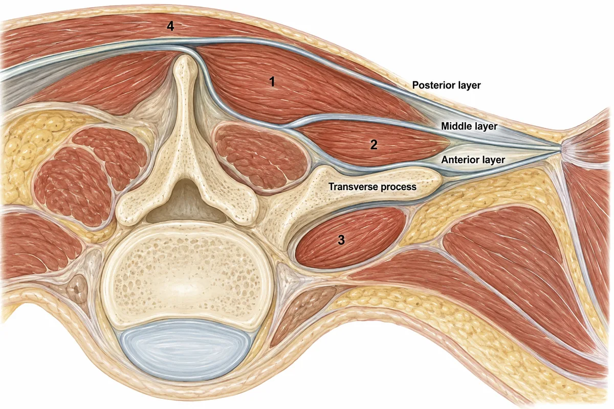
A transverse section of the posterior abdominal wall at the third lumbar vertebra is drawn in the figure with the fascial layers traced and four muscles numbered. Which muscle is number 2?
Look first: name the three layers of the fascia first; each muscle lies in the room between two of them.
 View larger ↗⤢ Tap to enlarge
- AQuadratus lumborum
- BPsoas major
- CErector spinae
- DLatissimus dorsi
Show the answer and explanation
Answer: A. Quadratus lumborum
Quadratus lumborum. The middle layer attaches to the tips of the transverse processes and the anterior layer to their front surfaces, so the room between them lies in front of the processes. Quadratus lumborum fills it, running from the iliac crest to the twelfth rib. Erector spinae sits in the room behind, between the posterior and middle layers.
Why the closest rival loses: psoas major (B) is muscle 3, in front of the anterior layer and not enclosed by it. Erector spinae (C) is muscle 1. Latissimus dorsi (D) is muscle 4, outside all three layers.
Recallab question 55903 · Anatomy › Abdomen and Pelvis · reference: Gray's Anatomy for Students
Recallab example 4 · the peritoneum
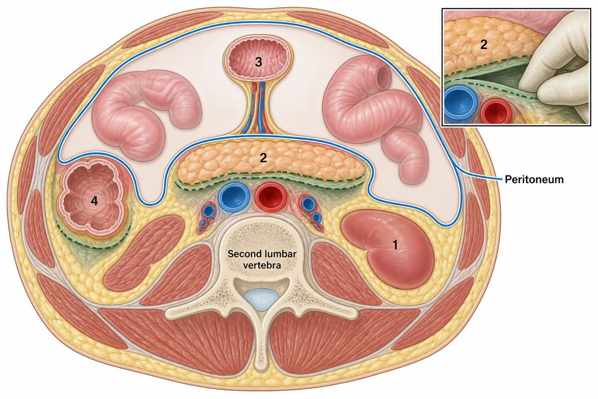
A transverse section of the abdomen is drawn in the figure with the peritoneum traced as a single continuous line and four organs numbered. Organ 2 came to lie behind the peritoneum only after it had first hung on a mesentery. What does that give the surgeon?
Look first: follow the blue peritoneal line and see which organs sit outside it, and which of those have a dotted line.
 View larger ↗⤢ Tap to enlarge
- AA bloodless fusion plane
- BNo plane at all
- CVessels to divide behind it
- DA mesentery to divide
Show the answer and explanation
Answer: A. A bloodless fusion plane
A bloodless fusion plane. An organ that was once on a mesentery and later fused to the back wall keeps the fusion plane, and that plane opens bloodlessly: it carries no vessels, so it separates with a finger rather than a knife. This is the reasoning behind rolling the colon, or the duodenum and pancreatic head, off the back wall.
Why the closest rival loses: no plane at all (B) describes organ 1, primarily retroperitoneal, which never had a mesentery to fuse. A mesentery to divide (D) describes organ 3, still suspended. The vessels (C) reach the organ from its old mesentery in front, not through the plane.
Recallab question 55105 · Anatomy › Abdomen and Pelvis · reference: Gray's Anatomy for Students
The neck and the brain
Recallab example 5 · the neck
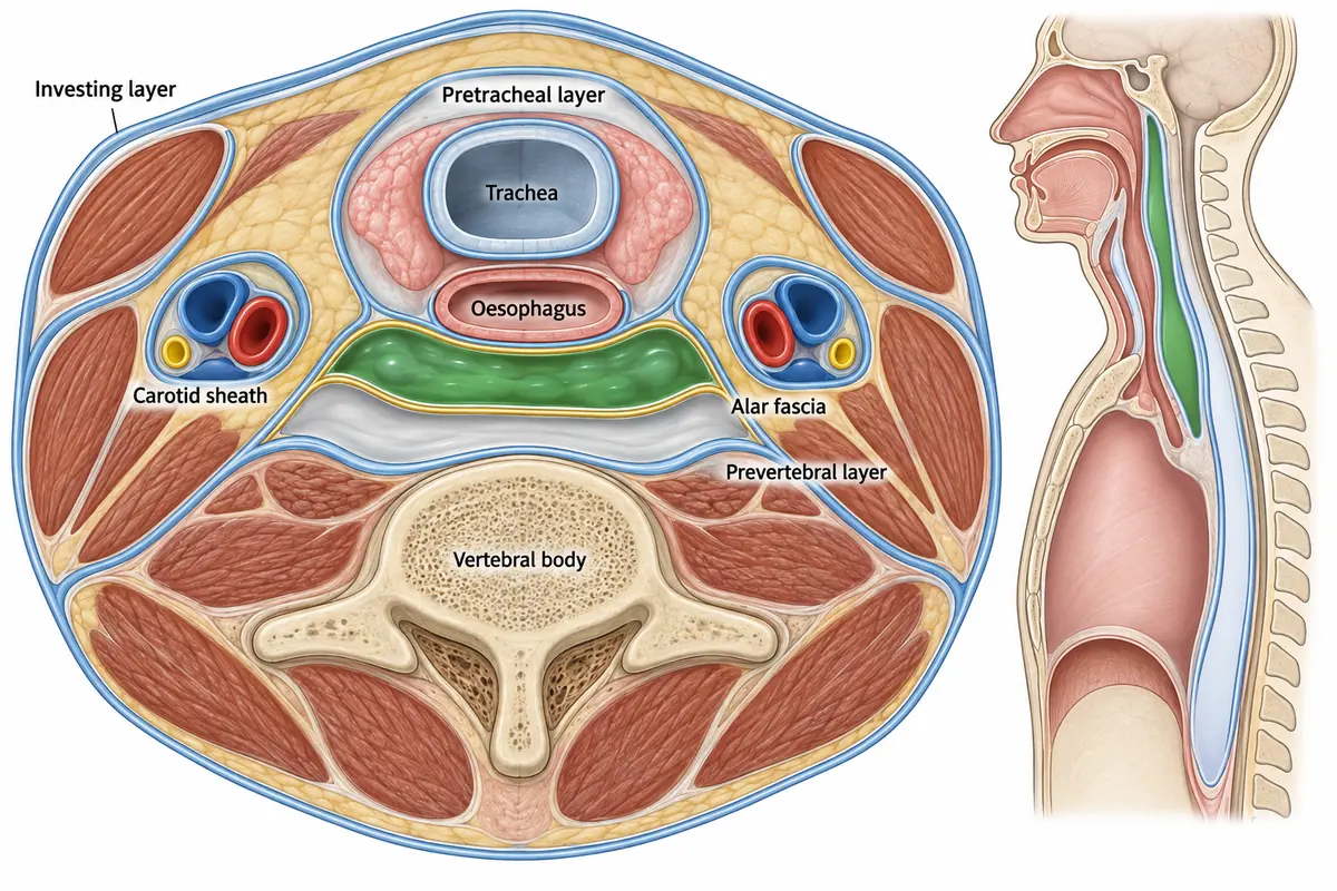
A transverse section of the neck is drawn in the figure with the fascial layers picked out and a collection shaded in one of the spaces. Where will that collection travel if it is not drained?
Look first: is the shaded space in front of the alar fascia or behind it?
 View larger ↗⤢ Tap to enlarge
- AInto the skull
- BInto the carotid sheath
- CTo the diaphragm
- DTo the upper mediastinum
Show the answer and explanation
Answer: D. To the upper mediastinum
To the upper mediastinum. The shaded collection lies in front of the thin alar fascia, so it is in the retropharyngeal space, which ends below where the alar fascia fuses with the pretracheal layer: at the upper mediastinum. The space behind the alar fascia has no such boundary and runs to the diaphragm, which is why it is called the danger space.
Why the closest rival loses: the diaphragm (C) is where the danger space behind the alar fascia ends, and that space is drawn empty here. The space is closed above at the skull base (A), and the carotid sheath (B) is a closed tube drawn separate from both.
Recallab question 55103 · Anatomy › Head and Neck · reference: Gray's Anatomy for Students
Recallab example 6 · the internal capsule
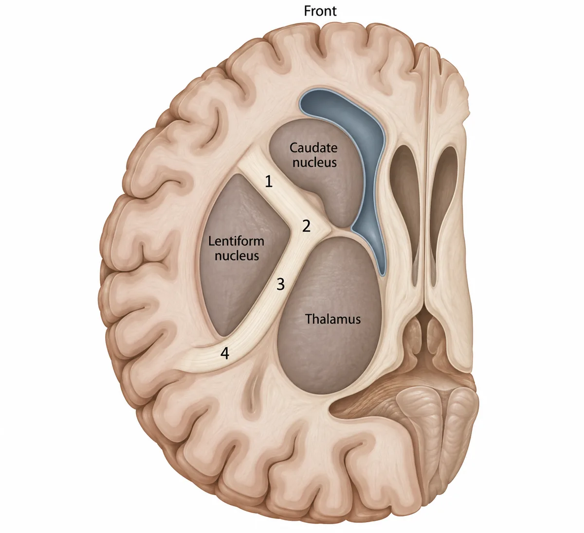
A small deep infarct causes weakness of the opposite face, arm and leg with no sensory loss and no loss of vision. A horizontal section of the cerebrum is drawn in the figure with four parts of the white matter numbered. Which numbered part holds the infarct?
Look first: name the nuclei on each side of the white matter V first: the thalamus and the lentiform nucleus.
 View larger ↗⤢ Tap to enlarge
- APart 4
- BPart 2
- CPart 1
- DPart 3
Show the answer and explanation
Answer: D. Part 3
Part 3, the posterior limb. The corticospinal fibres run between the thalamus and the lentiform nucleus, packed tightly behind the face fibres at the genu. One small infarct there weakens face, arm and leg together. The sensory and visual pathways lie further back, so they can be spared.
Why the closest rival loses: part 2 (B), the genu, carries the face fibres, so a lesion there alone would spare the arm and leg. Part 4 (A), behind the lentiform nucleus, carries the optic radiation, so a lesion there costs a field of vision. Part 1 (C), the anterior limb, carries frontopontine fibres, not the corticospinal tract.
Recallab question 55805 · Anatomy › Brain · reference: Snell's Clinical Anatomy by Regions
The one-minute routine
| If you see | Find first | It usually settles |
|---|---|---|
| A section through the arm | The two septa and the humerus | Which compartment, and so which nerve |
| A section across the wrist | The retinaculum and the trapezium | What is in the tunnel, and what is not |
| A section of the lumbar wall | The three fascial layers | The room each muscle lies in |
| A section of the abdomen | The peritoneal line | Which organs are retroperitoneal, and why |
| A section of the neck | The alar fascia | Retropharyngeal or danger space |
| A horizontal section of the brain | The thalamus and the lentiform nucleus | Which part of the internal capsule |
For other kinds of figure, see ten NEET PG image questions and six eye and ENT spotters. For the tools of the theatre, see surgical instruments and procedures. To practise the topics that matter most one at a time, see high impact topics for NEET PG.
For the bones themselves, see six NEET PG fracture and radiograph questions.
For pregnancy and the antenatal visit, see six NEET PG antenatal care questions.
For the localisation questions that follow the anatomy, see six NEET PG neurology localisation questions.
Why candidates use Recallab
- Built towards 22,000 — a batch at a time
- Illustrations drawn for the question
- Every option explained, not just the right one
- AI study tools that are live today
- Start free, today
Or see the plans, or try a timed mock section.
Register to practise NEET PG anatomy questionsFree account, no card needed. Practise with every option explained and a reference on every question.Drawn from questions #55004, #55902, #55903, #55105, #55103, #55805 in the Recallab bank, each with its own verified answer and current-edition reference. Last reviewed 5 October 2026.