
Recallab › Blog › Neurology localisation for NEET PG: six pictures and the question to ask first: where?
Neurology localisation for NEET PG: six pictures and the question to ask first: where?
Every neurology localisation question is the same question: where is the lesion? The findings are the clues, and each one belongs to a place.
Register to practise NEET PG neurology questions →Free account, no card needed.
These questions give you a picture of the brainstem, the cord or the limb and a set of findings, and ask where the damage is. The pictures below are illustrations drawn for the questions, not scans. Each is a real Recallab MentorQ, and each answer turns on one feature of the picture. Try each before you open the answer.
Before you localise: three questions
- Which findings are on which side? Put each sign on the side of the body and ask where its pathway crosses.
- Which pathways would one lesion have to cross? One small place that holds all the damaged pathways usually wins.
- What is spared? What is normal tells you where the lesion is not.
The brainstem
Recallab example 1 · the pons
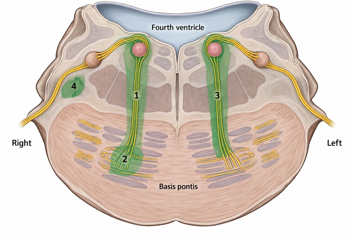
A man has weakness of the left arm and leg, weakness of the whole right side of his face, and cannot move either eye to the right. A transverse section of the lower pons is drawn in the figure with four lesion sites numbered. Which lesion explains all three findings?
Look first: put each finding on its side, then ask which lesion site lies where all three pathways run together.
 View larger ↗⤢ Tap to enlarge
- ALesion 3
- BLesion 1
- CLesion 2
- DLesion 4
Show the answer and explanation
Answer: B. Lesion 1
Lesion 1. The left limbs are weak, so the right corticospinal fibres are cut above their crossing. The whole right face is weak, which is a lower motor neurone palsy of the right facial nerve, whose fibres loop round the sixth nerve nucleus. That nucleus is a horizontal gaze centre: it holds motor neurones for the lateral rectus of its own side and interneurones for the opposite medial rectus. Destroying it paralyses gaze of both eyes towards the right, and only lesion 1 reaches from that nucleus down to the motor bundles in the basis.
Why the closest rival loses: lesion 2 (C) cuts the sixth nerve fibres after they leave the nucleus, which weakens only the right lateral rectus, so the left eye could still turn right. Lesion 3 (A) is on the left, which would weaken the right limbs and the left face, the mirror of this picture. Lesion 4 (D) is in the lateral tegmentum, which carries pain and temperature pathways: it causes sensory loss, not a gaze palsy or a hemiplegia. Nucleus lost, gaze lost; fibres cut, one muscle lost.
Recallab question 55803 · Anatomy › Brain · reference: Snell's Clinical Anatomy by Regions
Recallab example 2 · the medulla
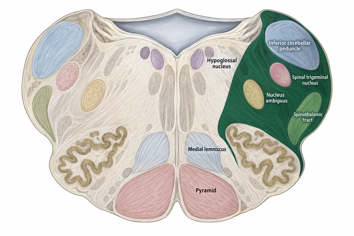
A transverse section of the medulla is drawn in the figure, with the territory lost after an arterial occlusion shaded. Which finding would NOT be expected?
Look first: list what lies inside the shaded wedge, then notice which structures lie outside it.
 View larger ↗⤢ Tap to enlarge
- AIpsilateral loss of facial pain sensation
- BContralateral arm and leg weakness
- CDysphagia and hoarseness
- DIpsilateral Horner syndrome
Show the answer and explanation
Answer: B. Contralateral arm and leg weakness
Contralateral arm and leg weakness. The shaded wedge is the lateral medulla, supplied by the vertebral or posterior inferior cerebellar artery. It holds the spinal trigeminal nucleus (facial pain loss on the same side), the spinothalamic tract (pain loss on the opposite side of the body), the nucleus ambiguus and the descending sympathetic fibres (dysphagia, hoarseness and Horner syndrome). The pyramid lies outside the wedge, so the limbs are not weak.
Why the closest rival loses: each of the other three findings is expected: facial pain loss (A) from the spinal trigeminal nucleus, dysphagia and hoarseness (C) from the nucleus ambiguus, and Horner syndrome (D) from the sympathetic fibres, all inside the wedge. Lateral medulla: crossed pain loss, no weakness.
Recallab question 55464 · Medicine › Neurology · reference: Harrison's Principles of Internal Medicine
The cerebellum
Recallab example 3 · the gait
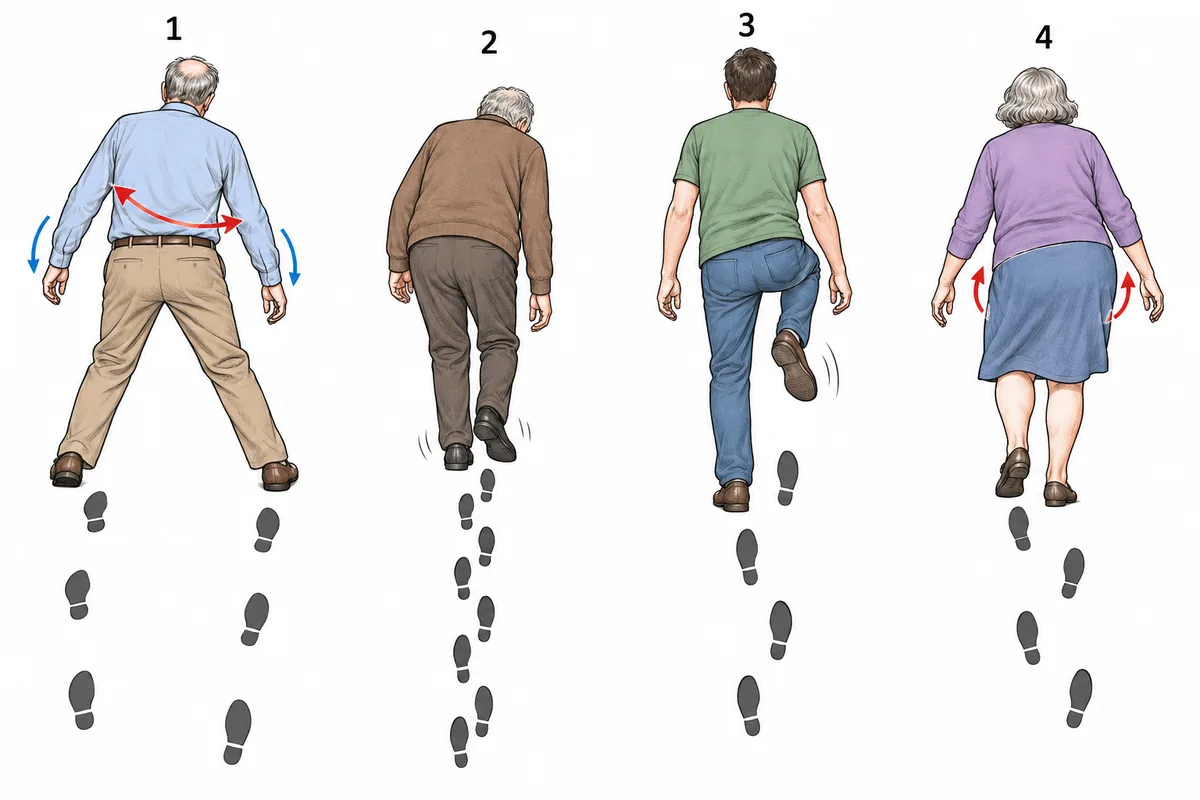
Four gaits are drawn in the figure. A man with long-standing heavy alcohol use walks with gait 1. Which part of the cerebellum is damaged?
Look first: describe the width of the feet and the sway of the trunk, then ask whether the arms are affected.
 View larger ↗⤢ Tap to enlarge
- ADentate nucleus
- BAnterior superior vermis
- CFlocculonodular lobe
- DLateral cerebellar hemisphere
Show the answer and explanation
Answer: B. Anterior superior vermis
The anterior superior vermis. Gait 1 is wide-based, with the trunk swaying and little incoordination of the arms, which is the pattern of vermis disease. Chronic alcohol use, with the thiamine deficiency that goes with it, damages this part selectively. A reminder that follows from the same story: thiamine is given before glucose, because a glucose load can use up the remaining thiamine and precipitate Wernicke encephalopathy.
Why the closest rival loses: the lateral cerebellar hemisphere (D) and the dentate nucleus (A) give limb ataxia, an intention tremor and dysmetria, not this trunk-dominant sway. The flocculonodular lobe (C) gives vertigo, nystagmus and instability even when sitting, typically from a midline tumour in a child. Vermis sways the trunk.
Recallab question 55671 · Medicine › Neurology · reference: Harrison's Principles of Internal Medicine
The cord, the root and the plexus
Recallab example 4 · the dorsal columns
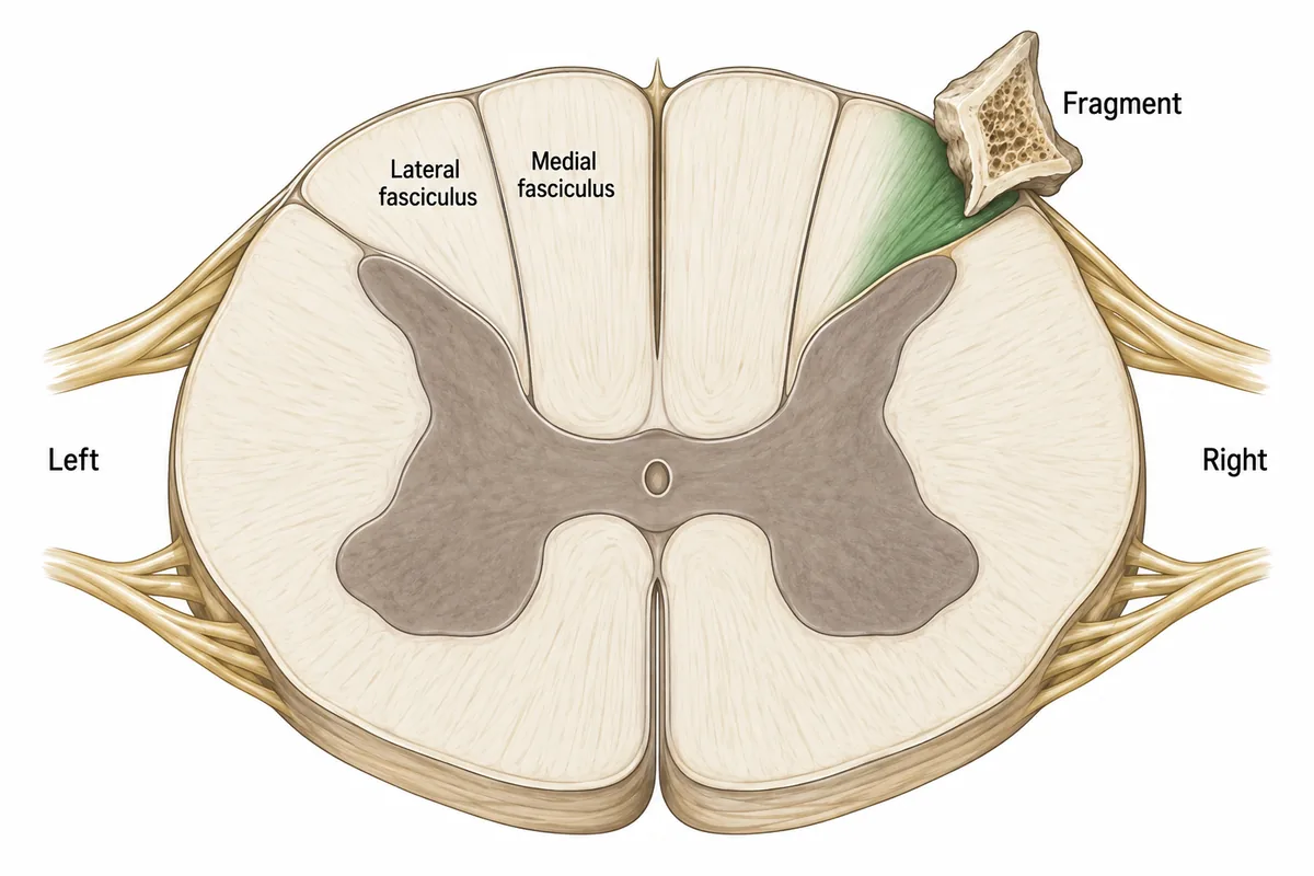
After a road accident, a bone fragment is found pressing on the back of the cervical spinal cord. A transverse section of the cord at that level is drawn in the figure with the fragment in place. Which loss is expected?
Look first: find the side of the fragment, then ask which fasciculus lies under it and what that fasciculus carries.
 View larger ↗⤢ Tap to enlarge
- ARight arm proprioception lost
- BRight leg proprioception lost
- CLeft arm pain sense lost
- DLeft arm proprioception lost
Show the answer and explanation
Answer: A. Right arm proprioception lost
Right arm proprioception lost. The dorsal columns are laminated: leg fibres lie medially in the fasciculus gracilis and arm fibres laterally in the fasciculus cuneatus. The fragment presses on the lateral fasciculus of the right dorsal column, which carries position and vibration sense from the upper limb. Dorsal column fibres ascend uncrossed and cross only in the medulla, so the loss is on the same side as the lesion.
Why the closest rival loses: right leg proprioception (B) is carried in the medial fasciculus, gracilis, which the fragment spares. Left arm proprioception (D) is wrong because the fibres have not yet crossed. Left arm pain sense (C) is carried by fibres that cross within a segment or two and ascend in the anterolateral cord, away from the fragment. Gracilis for the leg, cuneatus for the arm, and neither has crossed in the cord.
Recallab question 56108 · Physiology › Nervous System · reference: Guyton and Hall Textbook of Medical Physiology
Recallab example 5 · the cape
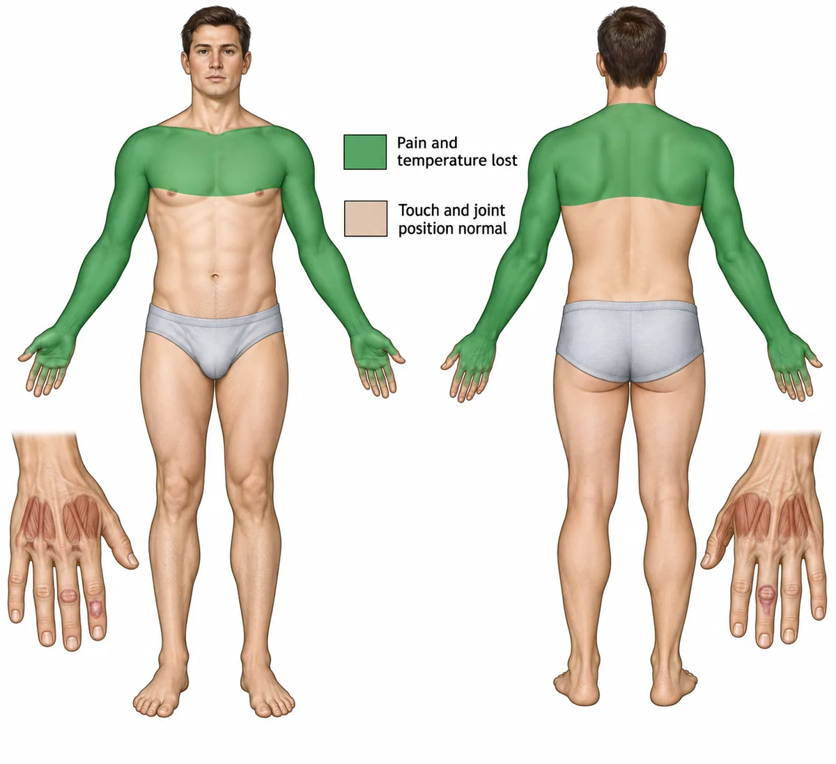
The sensory findings of a young man with painless burns on his hands are drawn in the figure as a body map. Which lesion explains them?
Look first: read the map for which sensations are lost and which are kept, and for the shape of the area.
 View larger ↗⤢ Tap to enlarge
- AA central cervical cord cavity
- BA cervical cord hemisection
- CPosterior column degeneration
- DBilateral carpal tunnel syndrome
Show the answer and explanation
Answer: A. A central cervical cord cavity
A central cervical cord cavity. Pain and temperature fibres cross just in front of the central canal, so a cavity there cuts them at every segment it spans. The loss is bilateral and cape-shaped over those segments, while the posterior columns lie behind and are spared, so touch and position stay normal. Spread into the anterior horns wastes the small hand muscles, and the hands, which no longer feel heat, are burned without pain: syringomyelia.
Why the closest rival loses: a hemisection (B) gives one-sided weakness and position loss with opposite-sided pain loss, not a symmetrical cape. Posterior column degeneration (C) loses position and vibration and spares pain, the reverse of this map. Bilateral carpal tunnel syndrome (D) numbs the palmar thumb and fingers and spares the forearms and shoulders. Cape of lost pain, touch intact: a syrinx.
Recallab question 55267 · Medicine › Neurology · reference: Harrison's Principles of Internal Medicine
Recallab example 6 · the arm
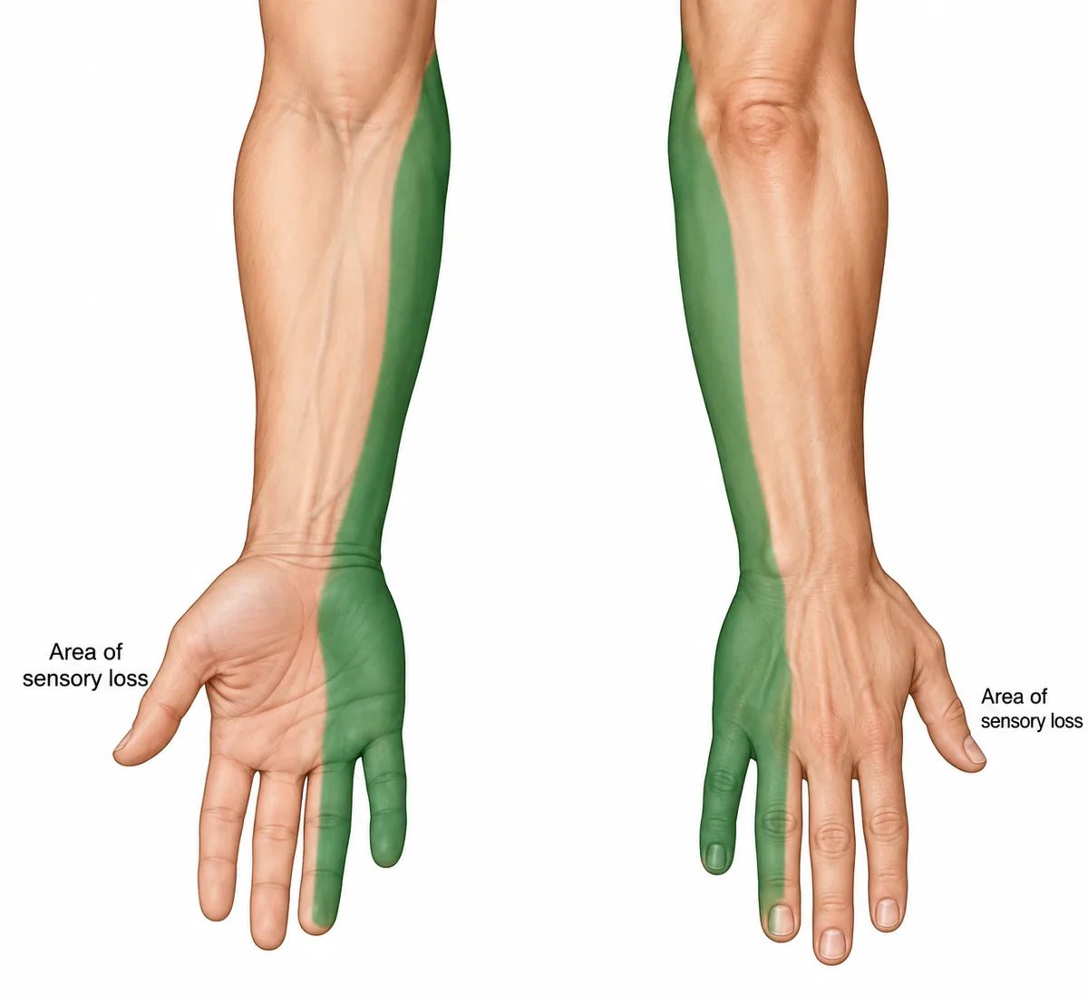
A woman has a weak hand. The area of sensory loss is shaded on the arm in the figure. Where is the lesion?
Look first: trace the edge of the shaded area and ask whether it stops at the wrist or carries on up the forearm.
 View larger ↗⤢ Tap to enlarge
- AThe ulnar nerve at the wrist
- BThe median nerve at the wrist
- CLower trunk or C8 and T1 roots
- DThe ulnar nerve at the elbow
Show the answer and explanation
Answer: C. Lower trunk or C8 and T1 roots
Lower trunk or C8 and T1 roots. The ulnar nerve supplies the little finger, half the ring finger and the ulnar palm, and it supplies no skin above the wrist at all. The medial forearm is supplied by the medial cutaneous nerve of the forearm, which arises from the medial cord, itself from C8 and T1. Loss in both territories therefore places the lesion at the roots or the lower trunk of the brachial plexus.
Why the closest rival loses: the ulnar nerve at the elbow (D) still supplies no forearm skin, so the medial forearm would be normal. The ulnar nerve at the wrist (A) would spare the back of the hand, which is shaded here. The median nerve at the wrist (B) supplies the thumb, index and middle fingers, all unshaded. Numbness above the wrist is never a hand nerve.
Recallab question 54770 · Medicine › Neurology · reference: Harrison's Principles of Internal Medicine
The one-minute routine
| If you see | Ask first | It usually settles |
|---|---|---|
| A section of the pons | Which findings are on which side | Nucleus or fibres |
| A section of the medulla with a wedge | What lies inside and what lies outside | Crossed pain loss, no weakness |
| Four gaits | Is the sway in the trunk or the limbs | Vermis or hemisphere |
| A cord section with a fragment | Which side, which fasciculus | What is lost, and on which side |
| A body map of lost pain | Which sensations are kept | A cavity in the central cord |
| A shaded arm | Does the area stop at the wrist | Nerve, or root and plexus |
For other kinds of figure, see ten NEET PG image questions and six eye and ENT spotters. For the decision that follows the picture, see next-best-step management questions.
Why candidates use Recallab
- Built towards 22,000 — a batch at a time
- Illustrations drawn for the question
- Every option explained, not just the right one
- AI study tools that are live today
- Start free, today
Or see the plans, or try a timed mock section.
Register to practise NEET PG neurology questionsFree account, no card needed. Practise with every option explained and a reference on every question.Drawn from questions #55803, #55464, #55671, #56108, #55267, #54770 in the Recallab bank, each with its own verified answer and current-edition reference. Last reviewed 10 October 2026.