
Recallab › Blog › Antenatal care for NEET PG: six pictures from the booking visit to the growth scan
Antenatal care for NEET PG: six pictures from the booking visit to the growth scan
Antenatal care questions test what a clinician does at the visit: what to advise, what to measure and when to act today rather than next week.
Register to practise NEET PG obstetrics questions →Free account, no card needed.
These questions show a picture from the antenatal clinic and ask for the advice, the instrument or the next step. The pictures below are illustrations drawn for the questions, not patient images. Each is a real Recallab MentorQ, and each answer turns on one feature of the drawing. Try each before you open the answer.
Before you answer an antenatal question: three questions
- What is the gestation? Many answers change with the week: when a sound can first be heard, when a measurement is reliable, when a symptom matters.
- Is this normal for her, or a change? A reading outside the range, or a fall from her own pattern, is the trigger.
- Is the action for today? Some findings are reassured and rechecked, others are assessed the same day.
Safe in pregnancy
Recallab example 1 · lying flat at term
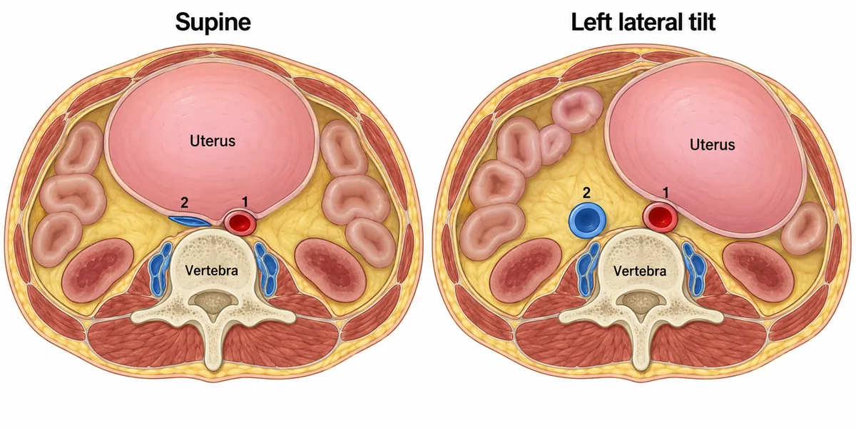
A woman at 36 weeks becomes pale, dizzy and hypotensive a few minutes after lying flat for an examination. Two transverse sections of her abdomen are drawn in the figure, one lying flat and one tilted, and vessel 2 is the one compressed. What follows from it?
Look first: name the two numbered vessels first, then ask what the uterus does to the one that is compressed.
 View larger ↗⤢ Tap to enlarge
- AA vagal bradycardia causes the faint
- BPlacental perfusion is unaffected
- CArterial inflow to the legs falls
- DVenous return and cardiac output fall
Show the answer and explanation
Answer: D. Venous return and cardiac output fall
Venous return and cardiac output fall. Vessel 2 is the inferior vena cava, the thin-walled vein to the right of the spine, so it flattens under the uterus when she lies flat. Less blood returns to the heart, so stroke volume and cardiac output fall. Most women compensate through paravertebral veins and a rise in tone; a few cannot. Left lateral tilt rolls the uterus off the cava and reverses the fall within minutes.
Why the closest rival loses: arterial inflow to the legs (C) is not the problem: vessel 1, the aorta, keeps its lumen because of its thick wall. A vagal bradycardia (A) is not the cause: the collapse is mechanical and recovers with tilt. The claim that placental perfusion is unaffected (B) is wrong: it falls too, so the fetal heart rate can drop. The same effect is why chest compressions in late pregnancy are given with the uterus displaced to the left.
Recallab question 55971 · Obstetrics › Physiological Changes of Pregnancy · reference: DC Dutta's Textbook of Obstetrics
Recallab example 2 · the seat belt
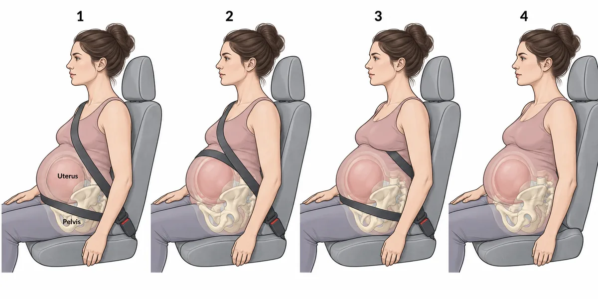
A woman at thirty weeks asks how she should wear a seat belt in a car. Four ways of wearing it are drawn in the figure. Which arrangement is correct?
Look first: follow each strap to see whether it crosses the bony pelvis or the uterus.
 View larger ↗⤢ Tap to enlarge
- ADrawing 3
- BDrawing 4
- CDrawing 1
- DDrawing 2
Show the answer and explanation
Answer: C. Drawing 1
Drawing 1. The lower strap goes under the bump and across the front of the bony pelvis, and the diagonal strap passes between the breasts and down the side of the abdomen. The bony pelvis can take a restraint load; the pregnant uterus cannot. A strap lying across the fundus presses the uterus against the spine on deceleration, and because the placenta does not stretch with the uterine wall, it shears away: abruption, the commonest cause of fetal death in a survivable crash.
Why the closest rival loses: drawing 2 (D) has its lower strap directly over the uterus, which delivers the crash load to the placental bed. Drawing 3 (A) has the diagonal strap tucked under the arm, which lets the upper body jack-knife over the lap strap. Drawing 4 (B), with no belt, is worse than any belt: being thrown forward is more dangerous than the strap. Under the bump, between the breasts.
Recallab question 58372 · Obstetrics › Antenatal Care · reference: DC Dutta's Textbook of Obstetrics
Listening and measuring
Recallab example 3 · hearing the heartbeat
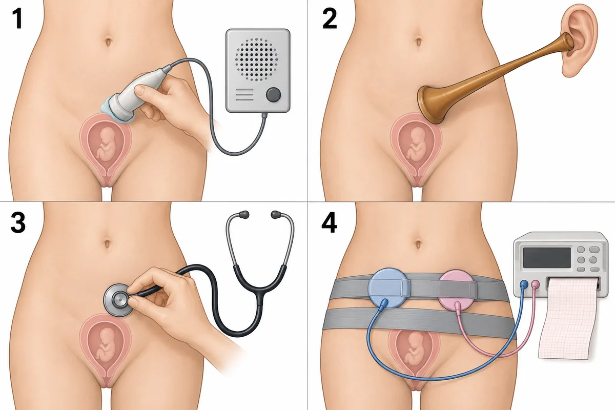
At her booking visit at 14 weeks, a woman asks to hear her baby's heartbeat. Four ways of listening over her lower abdomen are drawn in the figure. Which one will reliably pick up the heartbeat at this stage?
Look first: match each device to the gestation at which it first works.
 View larger ↗⤢ Tap to enlarge
- ADevice 3
- BDevice 1
- CDevice 4
- DDevice 2
Show the answer and explanation
Answer: B. Device 1
Device 1, a handheld Doppler. It turns the frequency shift from moving fetal blood into sound and detects the heart through the abdominal wall from about 10 to 12 weeks. At 14 weeks the heart sounds themselves are far too faint to hear directly. Transvaginal ultrasound shows the cardiac activity from about 6 weeks.
Why the closest rival loses: device 2 (D) is the Pinard stethoscope, which carries the heart sounds themselves and hears them only from about 20 weeks. Device 3 (A), an ordinary stethoscope, is even less suited, since direct listening works only later. Device 4 (C) is a cardiotocograph, which records the heart rate over time in late pregnancy and labour. Scan at 6, Doppler at 12, Pinard at 20.
Recallab question 58072 · Obstetrics › Diagnosis Of Pregnancy · reference: DC Dutta's Textbook of Obstetrics
Recallab example 4 · the fundal height
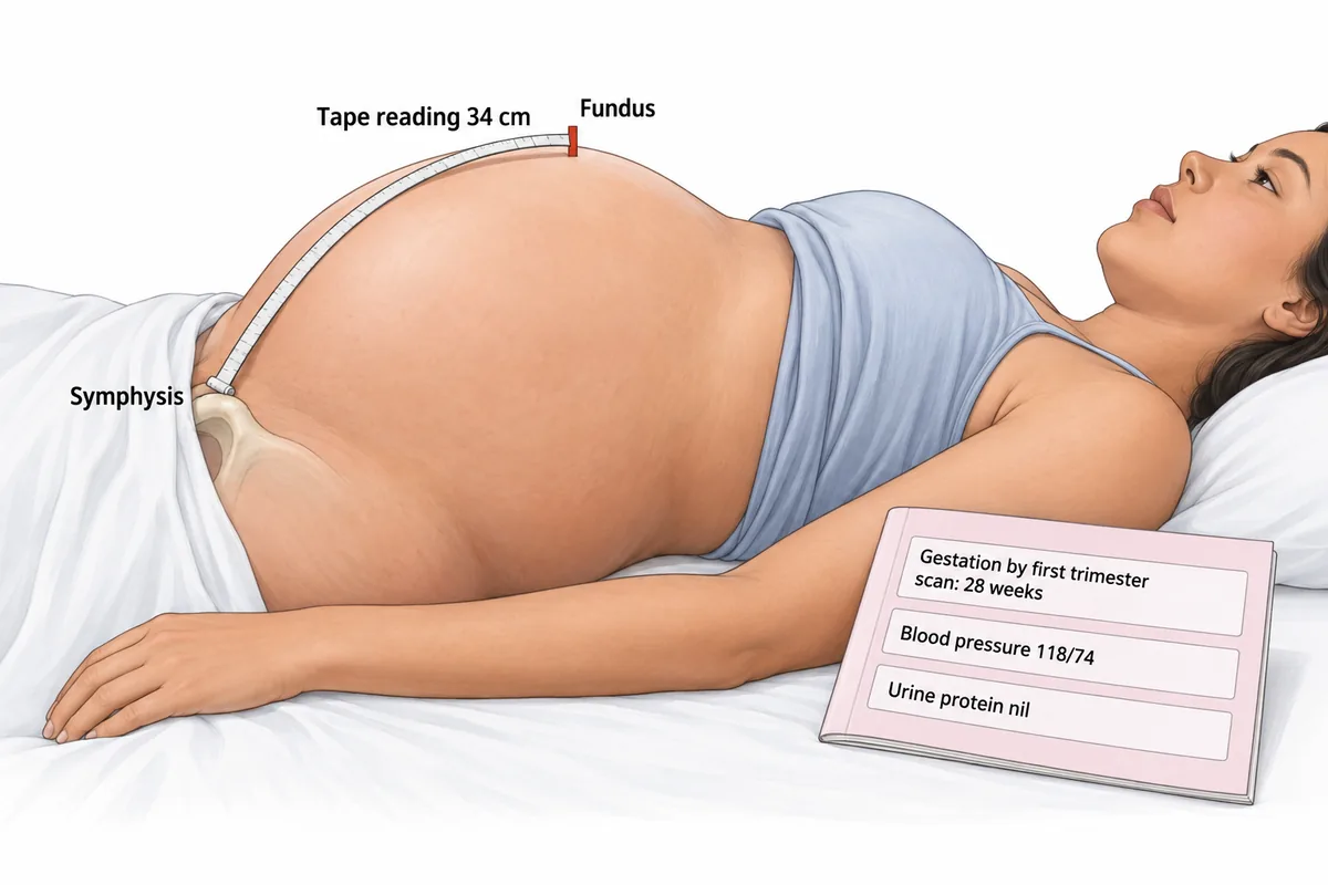
A woman's abdomen, measured with a tape from the symphysis, is drawn in the figure beside her Mother and Child Protection card. Her gestation was fixed by a first-trimester scan. What should be done next?
Look first: compare the tape reading with the gestation on the card, and ask how secure the dates are.
 View larger ↗⤢ Tap to enlarge
- AGlucose tolerance test
- BReassurance
- CRepeat the measurement in four weeks
- DUltrasound scan
Show the answer and explanation
Answer: D. Ultrasound scan
An ultrasound scan. After 24 weeks the height in centimetres should lie within about two to three of the gestation in weeks. Here the tape reads 34 cm at 28 weeks, six centimetres too large, and the dates are secure because an early scan fixed them. So something beyond a single normal fetus fills the uterus: twins, excess fluid, a large baby or a fibroid. One scan tells these apart, so it comes first.
Why the closest rival loses: repeating the measurement in four weeks (C) only delays finding the cause when the reading is well outside the accepted range. Reassurance (B) is wrong because the tape detects a uterus that is too large as well as too small. A glucose tolerance test (A) is considered once the scan shows which cause is present. Centimetres equal weeks, give or take three.
Recallab question 55773 · Obstetrics › Antenatal Care · reference: DC Dutta's Textbook of Obstetrics
Checking the baby
Recallab example 5 · the movement charts
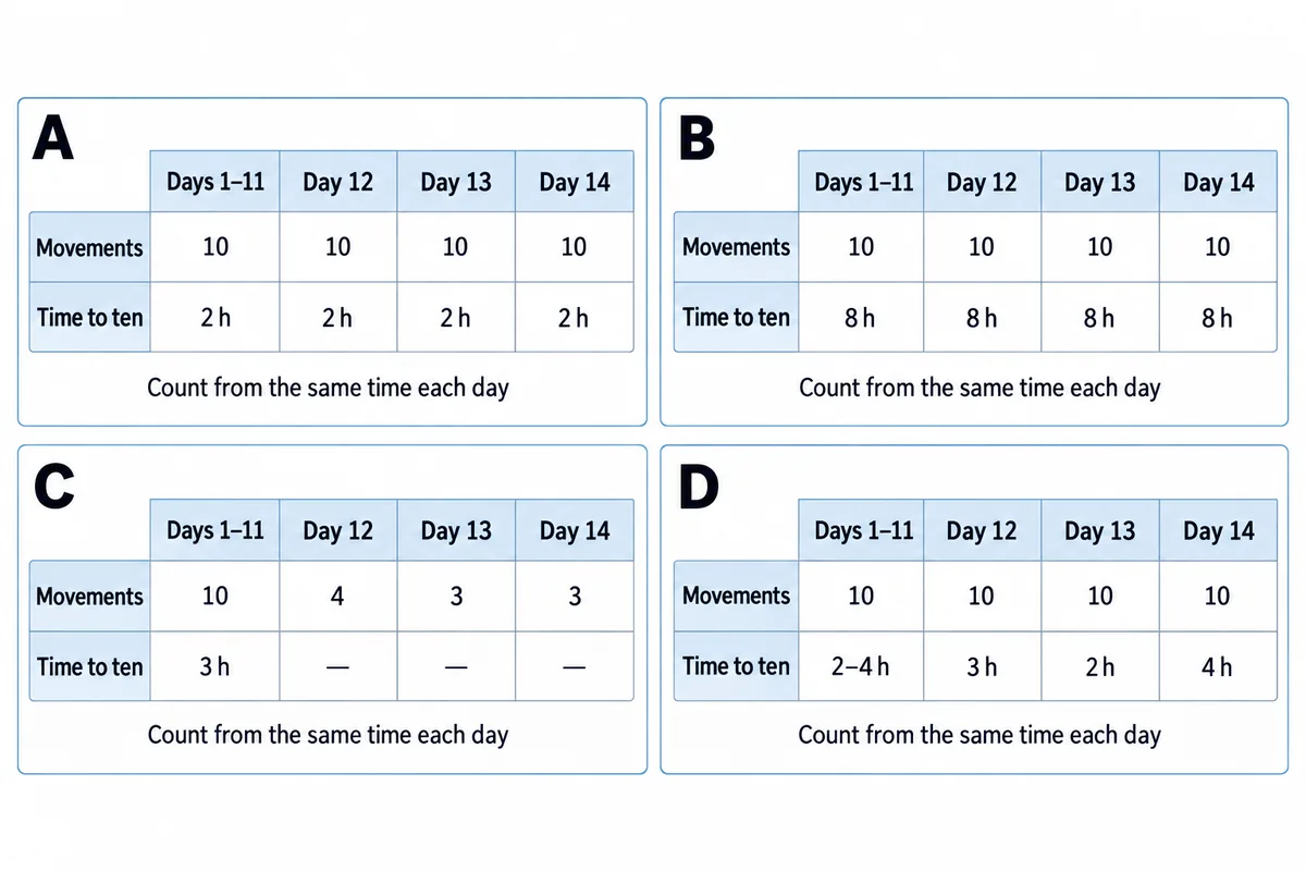
Four women at thirty-two weeks bring in the movement charts they have been keeping. The charts are drawn in the figure. Which woman must be assessed the same day?
Look first: compare each chart with that woman's own earlier days, not with the other charts.
 View larger ↗⤢ Tap to enlarge
- AWoman C
- BWoman A
- CWoman B
- DWoman D
Show the answer and explanation
Answer: A. Woman C
Woman C. Her chart records ten movements comfortably for eleven days and then falls short three days running. A fall from her own baseline matters more than any absolute number. A definite reduction is assessed the same day, with a history, a heart rate tracing and a scan for growth and liquor, because reduced movement precedes some stillbirths and is never left to the next clinic.
Why the closest rival loses: woman B (C) is slow, about eight hours to reach ten, but unchanged on every day, so it is her normal pattern. Woman A (B) reaches ten in about two hours every day. Woman D (D) shows ordinary day-to-day variation within a few hours and reaches ten every day. Her own pattern is the reference, and the visit is today.
Recallab question 58475 · Obstetrics › Antenatal assessment of Fetus · reference: DC Dutta's Textbook of Obstetrics
Recallab example 6 · the growth scan
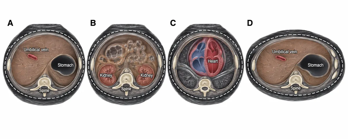
A growth scan is being done at thirty-four weeks and the sonographer has frozen four transverse sections of the fetal abdomen, lettered in the figure. Which section should the abdominal circumference be traced on?
Look first: name what each section shows, then ask which landmarks define the correct plane, and whether the outline is round.
 View larger ↗⤢ Tap to enlarge
- ASection B
- BSection C
- CSection D
- DSection A
Show the answer and explanation
Answer: D. Section A
Section A. The abdominal circumference is traced on the transverse section showing the stomach bubble and the short umbilical segment of the left portal vein within the liver, with the kidneys out of view. Those two landmarks fix the plane. A falsely large circumference conceals the growth restriction the scan was done to find.
Why the closest rival loses: section D (C) shows the same landmarks but its outline is an ellipse with three ribs on one side: an oblique cut, which inflates the measurement. Section B (A) is below the plane, because both kidneys are showing. Section C (B) is above it: the four-chambered heart marks the thorax. Stomach and vein in; kidneys and heart out; round, not oval.
Recallab question 57469 · Obstetrics › Antenatal assessment of Fetus · reference: DC Dutta's Textbook of Obstetrics
The one-minute routine
| If you see | Ask first | It usually settles |
|---|---|---|
| Sections lying flat and tilted | Which vessel is flattened | What falls, and the position to use |
| A seat belt on a pregnant woman | Does the strap cross the pelvis or the uterus | The safe arrangement |
| Devices over the lower abdomen | At what week does each first work | What can be heard at the booking visit |
| A tape and a card | Does the height match the weeks, and how secure are the dates | Reassure, or scan |
| Movement charts | Has this woman's own pattern changed | Same-day assessment or not |
| Sections of a fetal abdomen | Which landmarks are in view, and is it round | The right plane for the measurement |
For other kinds of figure, see ten NEET PG image questions and six eye and ENT spotters. For the decision that follows the picture, see next-best-step management questions.
Why candidates use Recallab
- Built towards 22,000 — a batch at a time
- Illustrations drawn for the question
- Every option explained, not just the right one
- AI study tools that are live today
- Start free, today
Or see the plans, or try a timed mock section.
Register to practise NEET PG obstetrics questionsFree account, no card needed. Practise with every option explained and a reference on every question.Drawn from questions #55971, #58372, #58072, #55773, #58475, #57469 in the Recallab bank, each with its own verified answer and current-edition reference. Last reviewed 8 October 2026.