
Recallab › Blog › Don't mix these up for NEET PG: extradural vs subdural haematoma
Don't mix these up for NEET PG: extradural vs subdural haematoma
A high-yield NEET PG pair. One layer separates them. Extradural blood sits outside the dura; subdural blood sits beneath it. Where the dura is fixed decides the shape, and the shape decides the answer.
Register to practise NEET PG neurosurgery questions →Free account, no card needed.
Extradural vs subdural haematoma: side by side
| Extradural haematoma | Subdural haematoma | |
|---|---|---|
| Location | Outside the dura | Beneath the dura |
| Bleeding vessel | Usually the middle meningeal artery | Bridging veins |
| Shape | Biconvex lens | Crescent |
| Sutures | Stops at sutures | Crosses sutures |
| Midline | Can cross | Limited by the falx |
This pair is one of the Don't mix these up questions in Recallab's NEET PG High yield screener.
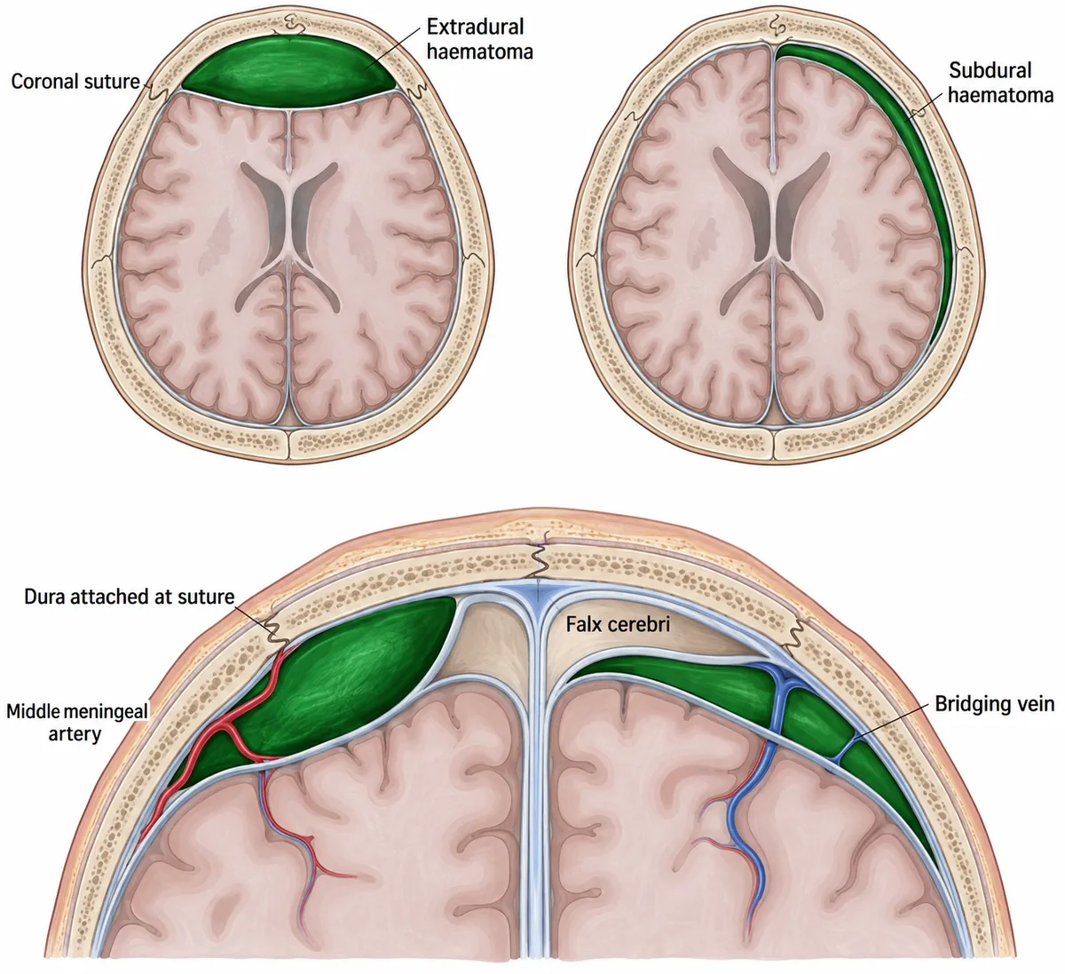
The dura is firmly attached to the skull at the sutures. Blood outside the dura is held between those attachments, so it pushes inward as a lens. Blood beneath the dura is not held by them, so it spreads along the brain as a crescent.
The one trap
The trap is confusing sutures with the midline. An extradural haematoma stops at sutures but can cross the midline. A subdural haematoma crosses sutures but cannot cross the midline, because the falx, a fold of dura, stops it there.
A lucid interval is classic for extradural haematoma, but it occurs in only a minority of cases. Its absence does not rule an extradural out.
Try it: say it once before the questions: lens stops at sutures; crescent stops at the falx.
Shape and vessel
Recallab example 1 · lucid, then deteriorating
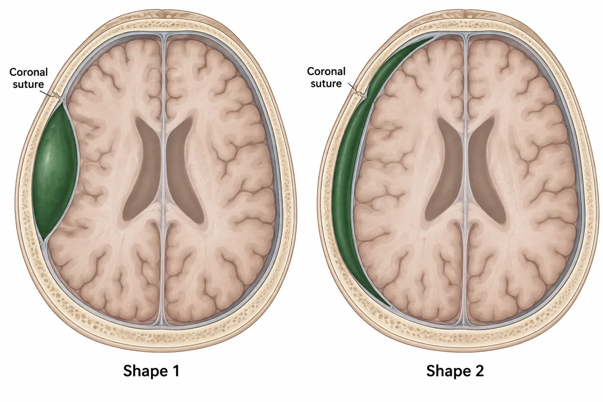
Two intracranial collections are drawn in the figure, with the skull sutures marked. A young man struck on the temple was briefly unconscious, then lucid, and is now deteriorating. Which shape fits, and what is bleeding?
Look first: find where each collection meets the coronal suture, then match the vessel to the space.
 View larger ↗⤢ Tap to enlarge
- AShape 1, a bridging vein
- BShape 1, middle meningeal artery
- CShape 2, middle meningeal artery
- DShape 2, middle cerebral artery
Show the answer and explanation
Answer: B. Shape 1, middle meningeal artery
B. Shape 1, middle meningeal artery. The dura is firmly attached to the inner skull at the suture lines. Blood collecting outside it is held between those attachments, so it pushes inward as a lens that stops at the suture. That is an extradural haematoma, and the vessel is the middle meningeal artery, which runs in a groove under the thin pterion. The blow causes a brief concussion, and the clot needs time to strip the dura from the bone and enlarge, which is why the patient is lucid before deteriorating.
Why the other options lose: Shape 1 with a bridging vein (A) has the right shape and the wrong vessel; bridging veins bleed into the subdural space. Shape 2 with the middle meningeal artery (C) pairs the subdural shape with the artery of the extradural space. Shape 2 with the middle cerebral artery (D) is wrong because that artery runs in the subarachnoid space; its bleeding goes into the subarachnoid space or the brain, not into either of these spaces.
Recallab question 54775 · Surgery › Cardiothoracic & Neurosurgery · reference: Bailey & Love's Short Practice of Surgery
Sutures and the midline
Recallab example 2 · a lens that crosses the midline
A head scan after a fall shows a dense biconvex collection that does not cross the coronal suture but does cross the midline. Which haematoma is this?
Look first: use the two boundaries: which layer is fixed at the sutures, and which one forms the falx?
- AExtradural haematoma
- BSubdural haematoma
- CIntracerebral haemorrhage
- DSubarachnoid haemorrhage
Show the answer and explanation
Answer: A. Extradural haematoma
 View larger ↗⤢ Tap to enlarge
A. Extradural haematoma. It usually follows a temporal fracture that tears the middle meningeal artery. Arterial pressure strips the dura from the bone, giving a biconvex shape, and the dura's attachment at the sutures stops it there. The falx limits a subdural collection at the midline, but an extradural collection lies outside the dura and can cross it.
Why the other options lose: Subdural haematoma (B) is crescentic, crosses sutures and is limited by the falx, the reverse of both features. Intracerebral haemorrhage (C) lies within the brain rather than against the skull. Subarachnoid haemorrhage (D) fills the sulci and basal cisterns rather than forming a lens.
Recallab question 51664 · Radiology › Neuroimaging & Head/Neck · reference: Bailey & Love's Short Practice of Surgery
The slow one
Recallab example 3 · three weeks of confusion on warfarin
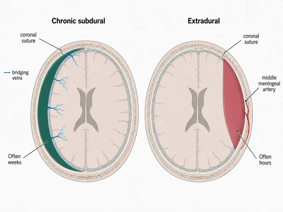
A man of 76 on warfarin has been increasingly confused and unsteady for three weeks, with a headache that varies through the day. He recalls no injury. CT shows a crescent-shaped hypodense collection over the left convexity that extends across the coronal suture. What is it?
Look first: read the shape and the suture first, then let the three weeks tell you how old the blood is.
- AAcute subarachnoid haemorrhage
- BChronic subdural haematoma
- CMiddle cerebral artery infarct
- DExtradural haematoma
Show the answer and explanation
Answer: B. Chronic subdural haematoma
 View larger ↗⤢ Tap to enlarge
B. Chronic subdural haematoma, from torn bridging veins. Brain atrophy in old age stretches those veins, so trivial force tears them, often too trivial to remember. Venous bleeding is slow, so symptoms take weeks to appear. The subdural space is not limited by the sutures, so the collection spreads as a crescent across them, and old blood has become hypodense, matching the three-week history. Anticoagulation, alcohol and previous falls are the usual risk factors.
Why the other options lose: Acute subarachnoid haemorrhage (A) gives hyperdense blood in the cisterns and sulci with a thunderclap onset, not a convexity crescent. A middle cerebral artery infarct (C) follows an arterial territory, with a focal deficit rather than fluctuating confusion. Extradural haematoma (D) is biconvex and stops at sutures; it is arterial and evolves over hours, not weeks.
Recallab question 50647 · Medicine › Neurology · reference: Harrison's Principles of Internal Medicine
Practise more Don’t mix these up stacks, where each pair of look-alikes is taught side by side. For more lesions placed by their anatomy, see neurology localisation.
Why candidates use Recallab
- Built towards 22,000 — a batch at a time
- Illustrations drawn for the question
- Every option explained, not just the right one
- AI study tools that are live today
- Start free, today
Or see the plans, or try a timed mock section.
Register to practise NEET PG neurosurgery questionsFree account, no card needed. Practise with every option explained and a reference on every question.Drawn from questions #54775, #51664, #50647 in the Recallab bank, each with its own verified answer and current-edition reference. Last reviewed 28 October 2026.