
Recallab › Blog › Blood films for NEET PG: six worked fields, from fragments to a crowded marrow
Blood films for NEET PG: six worked fields, from fragments to a crowded marrow
A blood film is solved by asking what the red cells have been through. Cells that were sheared, trimmed, starved or squeezed out each carry a different shape, and the shape tells you where to look next.
Register to practise NEET PG haematology questions →Free account, no card needed.
These six illustrated Recallab questions each put a film at the centre: three where red cells are being destroyed, and three where the film points to what is missing or what is crowding the marrow. Whether you are learning haematology in MBBS or revising for NEET PG or INI-CET, try each question before opening its worked answer.
Before you read a blood film: three questions
- What shape are the red cells? Fragments, small round dense cells, large oval cells and teardrops each point somewhere different.
- What else is in the field? Count the platelets, and look for immature cells, young blue-grey red cells and inclusions.
- What result goes with it? An antiglobulin test, the clotting times or one blood level usually separates two films that look alike.
Red cells being destroyed: sheared, coated or trimmed
Recallab example 1 · fragments and few platelets
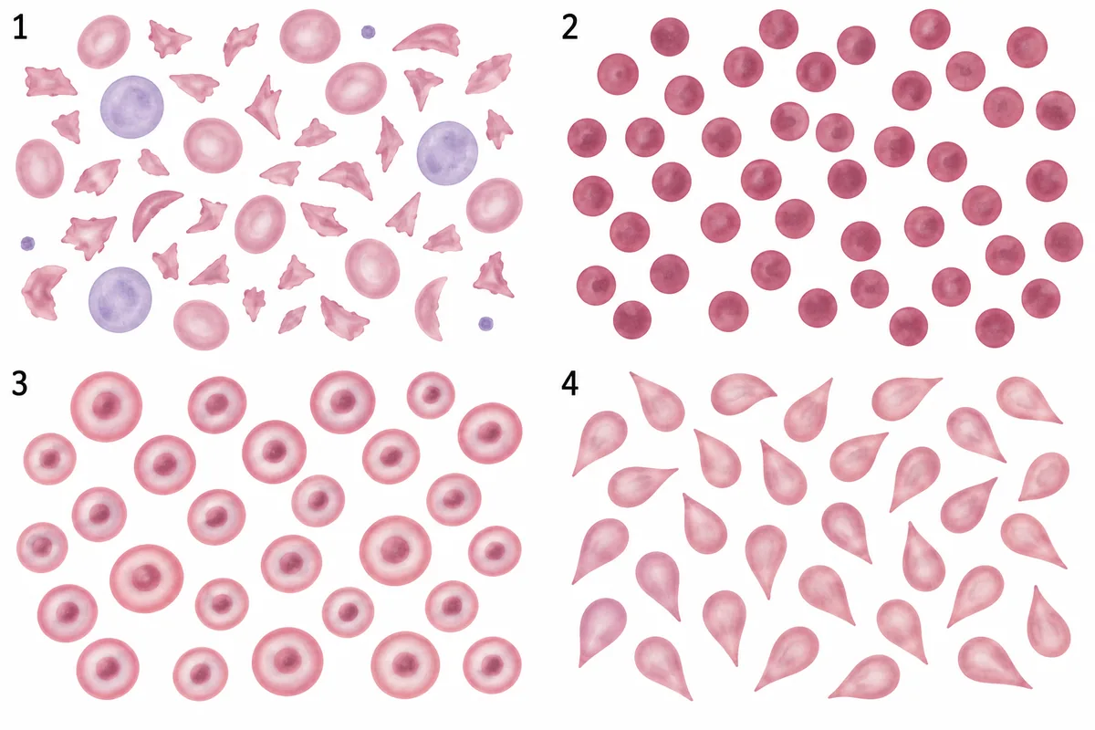
A woman has fever, confusion and falling platelets, with normal clotting times. Her blood film is drawn in the figure as panel 1, beside three other films. What are the cells in panel 1, and what process do they indicate? Four peripheral blood films, the first from a woman with fever and falling platelets.
Look first: name the shape in panel 1 first, then let the clotting times choose between the two causes of that shape.
 View larger ↗⤢ Tap to enlarge
- ASchistocytes, from microangiopathic haemolysis
- BSchistocytes, from disseminated intravascular coagulation
- CTarget cells, from thalassaemia
- DSpherocytes, from warm autoimmune haemolysis
Show the answer and explanation
Answer: A. Schistocytes, from microangiopathic haemolysis
A. Schistocytes, from microangiopathic haemolysis. Helmet cells, triangles and crescents are fragments of red cells, sheared as they pass platelet thrombi in small vessels. Fragments with a falling platelet count mean a thrombotic microangiopathy, and fever, confusion and normal clotting times point to thrombotic thrombocytopenic purpura. It is treated urgently with plasma exchange, a very low ADAMTS13 activity confirms it, and platelet transfusion is generally avoided.
Why the other options lose: Disseminated intravascular coagulation (B) also makes schistocytes, but it prolongs the clotting times, and here they are normal. Target cells (C) are panel 3. Spherocytes (D) are panel 2, small round dense cells with no pale centre. Fragments, low platelets, normal clotting: think of TTP.
Recallab question 55459 · Pathology › Blood RBC · reference: Robbins and Cotran Pathologic Basis of Disease
Recallab example 2 · spherocytes, a positive test and no platelets
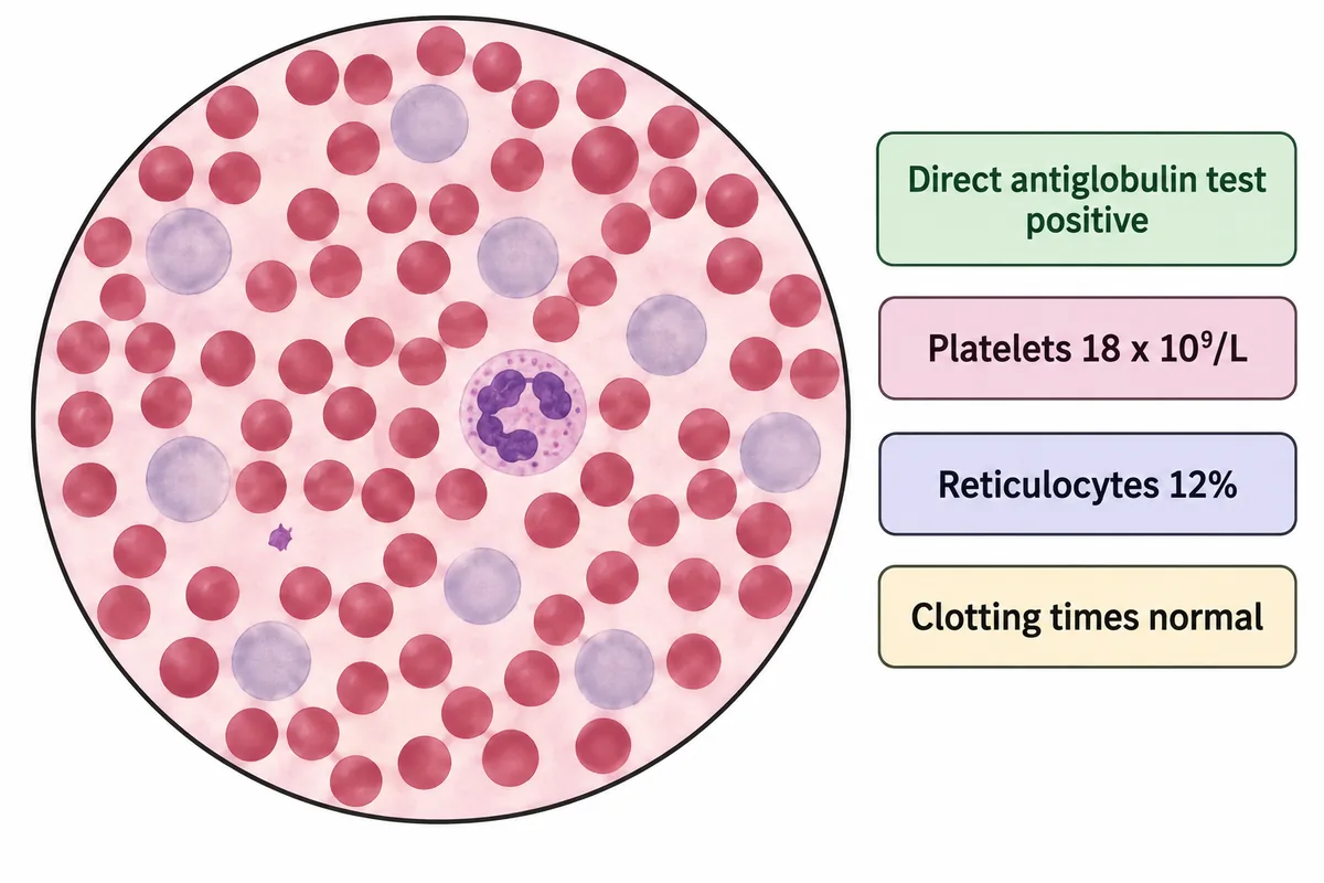
A woman of 26 has bruising, tiredness and yellow eyes. Her blood film and her results are drawn in the figure. What is the diagnosis? A peripheral blood film at high power beside four result tags.
Look first: name the small dense round cells, then count the platelets and read the antiglobulin test.
 View larger ↗⤢ Tap to enlarge
- AEvans syndrome
- BImmune thrombocytopenic purpura
- CThrombotic thrombocytopenic purpura
- DHereditary spherocytosis
Show the answer and explanation
Answer: A. Evans syndrome
A. Evans syndrome. The small dense round cells are spherocytes, trimmed by the spleen, and the larger blue-grey cells are reticulocytes, the marrow answering the haemolysis. A positive antiglobulin test for IgG shows that the red cells are coated with antibody, and the platelets are very low as well. Two antibody-mediated cytopenias together make Evans syndrome, and in a young woman lupus is the first underlying disease to look for.
Why the other options lose: Immune thrombocytopenic purpura (B) on its own gives no spherocytes, no jaundice and no positive antiglobulin test. Thrombotic thrombocytopenic purpura (C) gives fragments and a negative antiglobulin test, and this film has no fragments. Hereditary spherocytosis (D) looks the same on the film, but its antiglobulin test is negative and its platelets are normal. Spherocytes, a positive antiglobulin test and no platelets: Evans.
Recallab question 56857 · Medicine › Blood · reference: Harrison's Principles of Internal Medicine
Recallab example 3 · a leukaemia film that changed
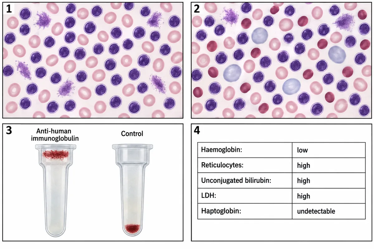
A man of 68 was found at a routine check to have a very high lymphocyte count and was left on observation. Three months later he is tired and pale. His two films, his antiglobulin card and his new results are drawn in the figure. What is the first treatment? Two blood films three months apart, an antiglobulin card and a panel of results.
Look first: compare the red cells, not the lymphocytes, between the two films, then read the card.
 View larger ↗⤢ Tap to enlarge
- ASplenectomy
- BOral iron
- CCorticosteroids
- DCombination chemotherapy
Show the answer and explanation
Answer: C. Corticosteroids
C. Corticosteroids. The lymphocytes are unchanged; what is new is in the red cells. The second film adds spherocytes and young blue-grey cells, the reticulocytes are high, the bilirubin and LDH are raised and the haptoglobin has gone, so the marrow is working hard against haemolysis rather than failing. The card shows immunoglobulin on the red cells, so the haemolysis is autoimmune, a recognised complication of chronic lymphocytic leukaemia, and it is treated first with steroids.
Why the other options lose: Splenectomy (A) is kept for haemolysis that fails steroids and later treatments. Oral iron (B) is for iron lack, which gives small pale cells, not spherocytes with a positive card. Combination chemotherapy (D) answers marrow replacement, which gives a low reticulocyte count and a negative antiglobulin test. An anaemia in chronic lymphocytic leukaemia is not automatically progression.
Recallab question 57520 · Pathology › Blood WBC & Lymphoma · reference: Robbins and Cotran Pathologic Basis of Disease
What is missing, and what is crowding the marrow
Recallab example 4 · a megaloblastic film after surgery
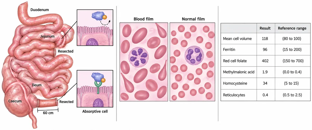
A woman of 34 has had two operations on her small bowel for Crohn disease and is tired and breathless on exertion. Her operative diagram, her blood film and her results are drawn together in the figure. Which single result separates the deficiency behind her film from its alternative? A small bowel diagram after two resections, the blood film, and a panel of results with their reference ranges.
Look first: see which length of bowel has gone, then find the one result that only one of the two vitamins can raise.
 View larger ↗⤢ Tap to enlarge
- ARaised homocysteine
- BHypersegmented neutrophils
- CRaised methylmalonic acid
- DRaised mean cell volume
Show the answer and explanation
Answer: C. Raised methylmalonic acid
C. Raised methylmalonic acid. Large oval red cells with a six-lobed neutrophil make a megaloblastic film, and the folate and ferritin are normal. The terminal ileum is the only place where vitamin B12 bound to intrinsic factor is absorbed, and that segment has been removed. Methylmalonyl-CoA mutase needs vitamin B12, so its substrate builds up only when vitamin B12 is lacking, which separates it from folate deficiency.
Why the other options lose: Raised homocysteine (A), hypersegmented neutrophils (B) and a raised mean cell volume (D) all appear in either deficiency, so none of them can separate the two. Methylmalonic acid is the tie-breaker: homocysteine rises in both, and only vitamin B12 deficiency raises the acid. After an ileal resection the replacement has to be by injection, for life.
Recallab question 57349 · Medicine › Blood · reference: Harrison's Principles of Internal Medicine
Recallab example 5 · golf-ball stippling
A blood film stained with brilliant cresyl blue is drawn in the figure in three panels. A man of Southeast Asian descent has a lifelong moderate microcytic haemolytic anaemia and splenomegaly. Which inclusion will his film show? A supravitally stained blood film drawn in three panels showing different red cell inclusions.
Look first: decide whether each inclusion is spread evenly through the cell or sits as one or two lumps at the edge.
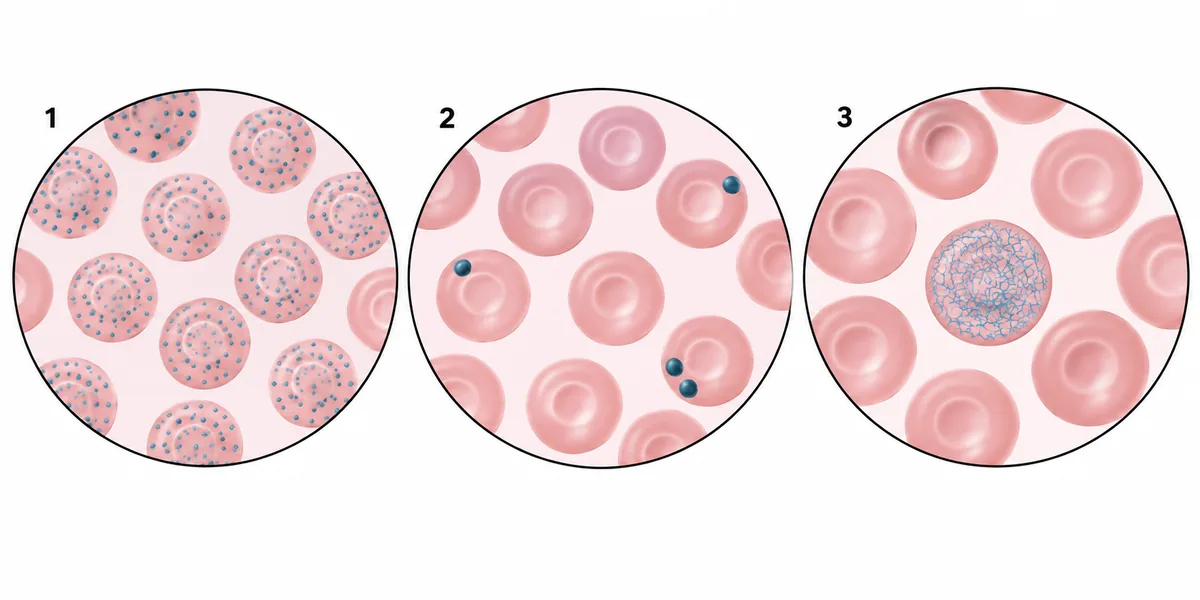 View larger ↗⤢ Tap to enlarge
- AHeinz bodies
- BReticulocytes
- CPappenheimer bodies
- DHaemoglobin H inclusions
Show the answer and explanation
Answer: D. Haemoglobin H inclusions
D. Haemoglobin H inclusions. Losing three of the four alpha-globin genes leaves unpaired beta chains, which form beta-4 tetramers, haemoglobin H, and precipitate inside the red cell. Supravital staining shows the precipitate as fine, even golf-ball stippling, as in panel 1. A lifelong microcytic haemolytic anaemia with splenomegaly in a man of Southeast Asian descent fits this alpha thalassaemia. Losing all four genes causes haemoglobin Bart's hydrops fetalis instead.
Why the other options lose: Heinz bodies (A) are the few large eccentric inclusions of panel 2, seen in G6PD deficiency after an oxidant trigger rather than lifelong. Reticulocytes (B) are panel 3, the normal RNA mesh of a young red cell. Pappenheimer bodies (C) are iron granules shown by Perls stain, not fine even stippling. Fine even stippling: haemoglobin H; eccentric lumps: Heinz bodies.
Recallab question 55324 · Pathology › Haematology: RBC · reference: Robbins and Cotran Pathologic Basis of Disease
Recallab example 6 · two immature lines at once
Four anaemic patients each had a blood film made, and the four fields are drawn in the figure. Which field says that the marrow space itself is being taken over? Four blood film fields from four anaemic patients.
Look first: look for cells that should still be in the marrow, and count how many cell lines they come from.
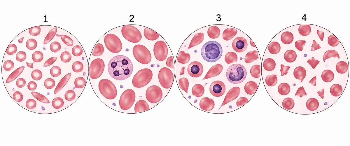 View larger ↗⤢ Tap to enlarge
- AField 4
- BField 1
- CField 2
- DField 3
Show the answer and explanation
Answer: D. Field 3
D. Field 3. Nucleated red cells and immature granulocytes in the blood together, with teardrop cells, make a leucoerythroblastic film. Immature cells of two lines at once are being pushed out of a marrow that is being crowded, by fibrosis, secondary deposits or a marrow tumour, and teardrops form as cells squeeze through the distorted marrow. The next step is to examine the marrow, and an aspirate may yield nothing.
Why the other options lose: Field 4 (A) shows fragments and a helmet cell with few platelets: red cells sheared in the circulation, not a crowded marrow. Field 1 (B) is the iron-lack film of small pale cells and pencil forms. Field 2 (C) is the megaloblastic film of large oval cells and a six-lobed neutrophil. Two immature lines at once: look in the marrow, not in the diet.
Recallab question 56221 · Pathology › Blood RBC · reference: Robbins and Cotran Pathologic Basis of Disease
The one-minute routine
| If the film shows | Ask first | It settles |
|---|---|---|
| Fragments with few platelets | Are the clotting times normal? | Normal: thrombotic microangiopathy such as TTP; prolonged: DIC |
| Spherocytes with few platelets | Is the antiglobulin test positive? | Positive: Evans syndrome; then look for lupus |
| Spherocytes appearing in a known leukaemia | Are the reticulocytes up and the card positive? | Autoimmune haemolysis: steroids first, not chemotherapy |
| Large oval cells and a six-lobed neutrophil | Is the methylmalonic acid raised? | Raised: vitamin B12 deficiency, not folate |
| Inclusions on a supravital stain | Fine and even, or one or two lumps at the edge? | Even stippling: haemoglobin H; eccentric lumps: Heinz bodies |
| Nucleated red cells and immature white cells | Are two immature lines out at once, with teardrops? | A crowded marrow: examine the marrow next |
Practise red cell questions, white cell and lymphoma questions and platelets and coagulation, and see more stained cells in microbiology and pathology spotters.
Why candidates use Recallab
- Built towards 22,000 — a batch at a time
- Illustrations drawn for the question
- Every option explained, not just the right one
- AI study tools that are live today
- Start free, today
Or see the plans, or try a timed mock section.
Register to practise NEET PG haematology questionsFree account, no card needed. Practise with every option explained and a reference on every question.Drawn from questions #55459, #56857, #57520, #57349, #55324, #56221 in the Recallab bank, each with its own verified answer and current-edition reference. Last reviewed 22 October 2026.