
Recallab › Blog › Microbiology and pathology spotters for NEET PG: six slides and where to look first
Microbiology and pathology spotters for NEET PG: six slides and where to look first
A microbiology or pathology spotter is a slide and a name. The marks go to the candidate who knows which one feature on the slide to look at first.
Register to practise NEET PG microbiology and pathology questions →Free account, no card needed.
The slides repeat from year to year: a Gram film, a yeast test, a stool cyst, a blood film, one odd cell in a lymph node. Most of what is drawn is background, and one detail settles the answer. Below are six real Recallab MentorQs, three from microbiology and three from pathology, each built on a figure. Try each before you open the answer.
Before you name anything: three questions
- What is the preparation? A Gram stain, a wet mount, a blood film or a tissue section. That limits the answers straight away.
- What is the background, and what is not? Normal cells are there for scale. Look for the one thing that should not be there.
- What does the question want? An organism, a disease, or which one of several panels.
Microbiology
Recallab example 1 · Gram film of CSF
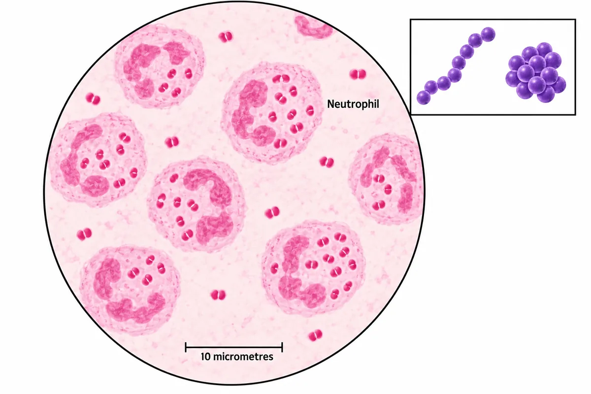
A Gram film of cerebrospinal fluid is drawn in the figure. Which organism does the appearance point to?
Look first: decide the colour before the shape: purple or pink?
 View larger ↗⤢ Tap to enlarge
- AStreptococcus pneumoniae
- BHaemophilus influenzae
- CStaphylococcus aureus
- DNeisseria meningitidis
Show the answer and explanation
Answer: D. Neisseria meningitidis
Rule: colour first, then shape, then where the organism lies. The cocci take the pink counterstain, so they are Gram negative. They sit in pairs with flattened facing sides, the coffee-bean shape. Many lie inside neutrophils. Cerebrospinal fluid is sterile, so organisms taken up by the patient's own cells point to true infection, not contamination.
Why the closest rival loses: the pneumococcus (A) is also a diplococcus, but it is Gram positive and lancet-shaped, so it would be purple. Haemophilus (B) is a small Gram-negative coccobacillus, not paired cocci. Staphylococci (C) are purple clusters, as the inset shows.
Recallab question 55022 · Microbiology › General Microbiology · reference: Ananthanarayan and Paniker's Textbook of Microbiology
Recallab example 2 · germ tube test
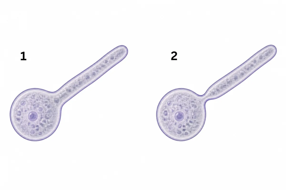
Two yeast isolates have been incubated in serum for two hours and examined, as in the figure. Which one is Candida albicans?
Look first: look only at where the filament leaves the cell.
 View larger ↗⤢ Tap to enlarge
- ANeither panel
- BPanel 1
- CBoth panels
- DPanel 2
Show the answer and explanation
Answer: B. Panel 1
Rule: a germ tube has no constriction where it leaves the cell. It is the start of a true hypha, continuous with the mother cell, so there is no cross wall and no narrowing at the junction. That is panel 1, and it identifies Candida albicans. The standard test is serum at 37 degrees for two to three hours.
Why the closest rival loses: panel 2 (D) is pinched at the junction. That is a pseudohypha, a chain of budding cells that never separated, and several Candida species make them. The constriction is the whole point of the test, so both panels (C) cannot count. Read it after three hours and other species produce filaments too.
Recallab question 54723 · Microbiology › Mycology · reference: Jawetz, Melnick & Adelberg's Medical Microbiology
Recallab example 3 · stool cysts
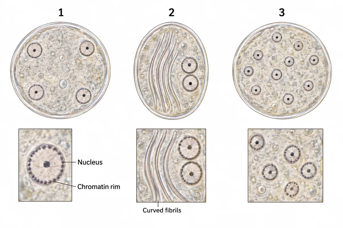
Three cysts from the same stool sample are drawn in the figure with their nuclei enlarged in a side panel. Which cyst is a harmless commensal that should not be treated, even though it resembles the other two on a quick look?
Look first: count the nuclei in each cyst, then look for anything that is not a nucleus.
 View larger ↗⤢ Tap to enlarge
- ACyst 1
- BCyst 3
- CCyst 2
- DCysts 2 and 3
Show the answer and explanation
Answer: B. Cyst 3
Rule: count the nuclei. Cyst 3 has eight or more, scattered: Entamoeba coli, a commensal that is not treated. Cyst 1 has exactly four, each with a small central karyosome: the mature Entamoeba histolytica cyst. Cyst 2's curved fibrils are the axonemes of Giardia lamblia.
Why the closest rival loses: cyst 1 (A) looks most like cyst 3 at a glance, but four nuclei with a central karyosome mark Entamoeba histolytica, a pathogen that is treated. Cyst 2 (C) is Giardia, a real cause of diarrhoea, so cysts 2 and 3 together (D) is wrong too.
Recallab question 56995 · Microbiology › Parasitology · reference: Ananthanarayan and Paniker's Textbook of Microbiology
Pathology
Recallab example 4 · blood films
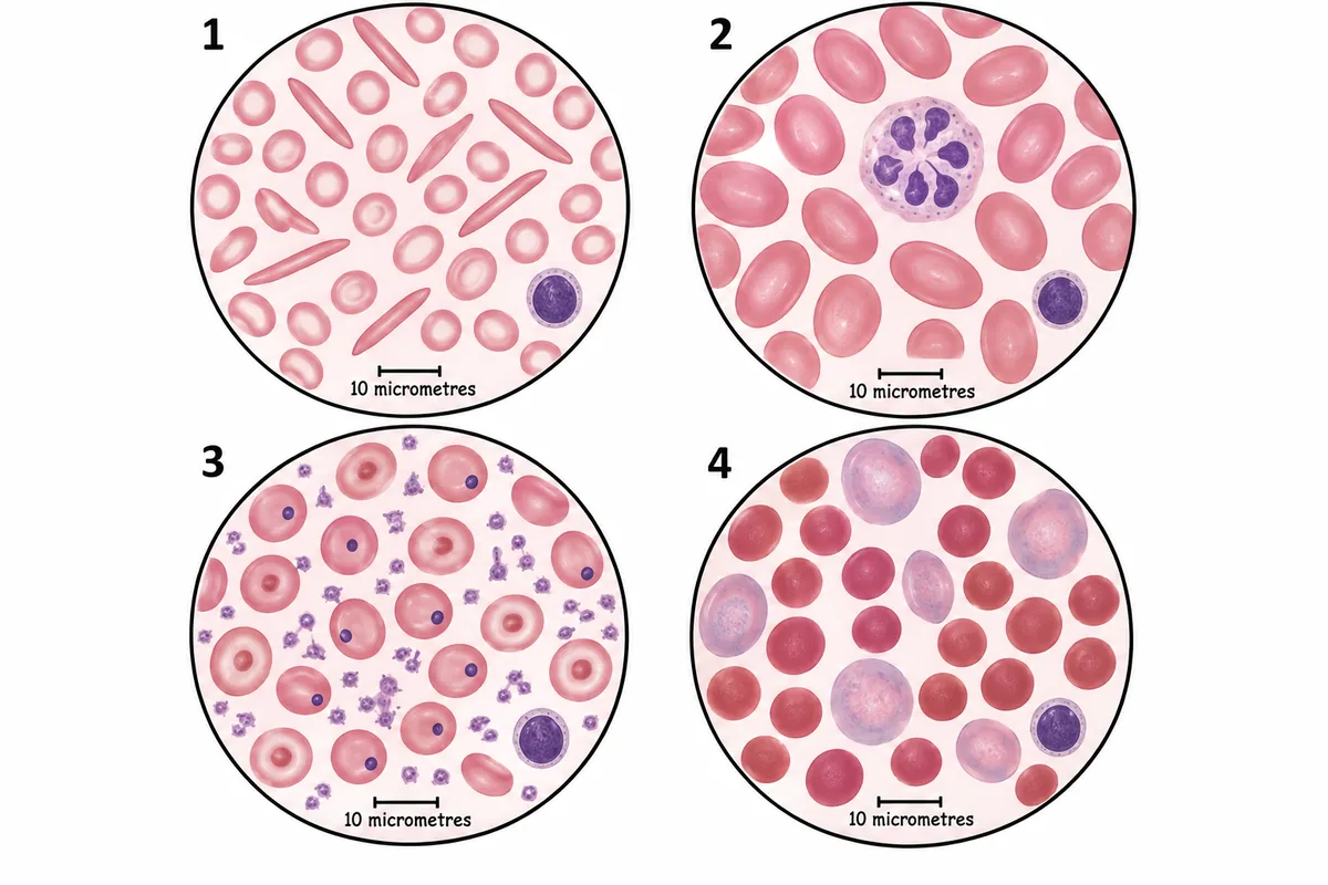
Four blood films are drawn in the figure at one magnification. Which film belongs to a patient whose spleen has been removed?
Look first: look inside the red cells and at the background, not at their size.
 View larger ↗⤢ Tap to enlarge
- AField 3
- BField 2
- CField 4
- DField 1
Show the answer and explanation
Answer: A. Field 3
Rule: after splenectomy, look for what the spleen would have removed. Film 3 has Howell-Jolly bodies, the single dark dots that are remnants of nucleus the spleen normally pits out. It also has target cells, with surplus membrane no longer trimmed, and a raised platelet count, because the spleen no longer pools them. Functional asplenia, as in sickle cell disease, gives the same picture.
Why the closest rival loses: the spherocytes of film 4 (C) are also a spleen story, but in hereditary spherocytosis the spleen is present and is doing the damage. Pencil cells (D) belong to iron deficiency, and the six-lobed neutrophil of film 2 (B) to megaloblastic anaemia.
Recallab question 55055 · Pathology › Haematology: RBC · reference: Robbins, Cotran & Kumar Pathologic Basis of Disease
Recallab example 5 · neutrophil nuclei
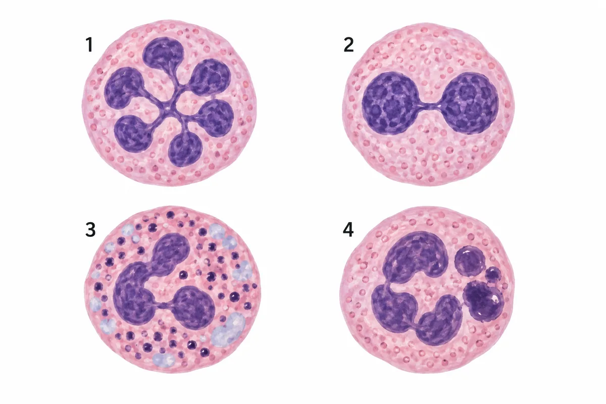
Four neutrophils from peripheral blood films are drawn in the figure. Which shows the Pelger-Huet anomaly?
Look first: count the lobes, then check the cytoplasm of the cells whose nucleus looks normal.
 View larger ↗⤢ Tap to enlarge
- ACell 4
- BCell 1
- CCell 3
- DCell 2
Show the answer and explanation
Answer: D. Cell 2
Rule: two lobes joined by a bridge, like spectacles, is Pelger-Huet. It is a benign autosomal dominant failure of nuclear lobing, from a lamin B receptor mutation, and the cells work normally. It matters because it can be mistaken for a left shift. The same nucleus appearing in an adult suggests myelodysplasia.
Why the closest rival loses: cell 1 (B) is the opposite fault: six lobes is hypersegmentation, a sign of megaloblastic anaemia. Cell 3 (C) has toxic granulation and Dohle bodies, the change of infection. Cell 4 (A) has the giant granules of Chediak-Higashi syndrome. Both have a normal nucleus.
Recallab question 55258 · Pathology › Haematology: WBC & Lymphoma · reference: Robbins, Cotran & Kumar Pathologic Basis of Disease
Recallab example 6 · Hodgkin cells
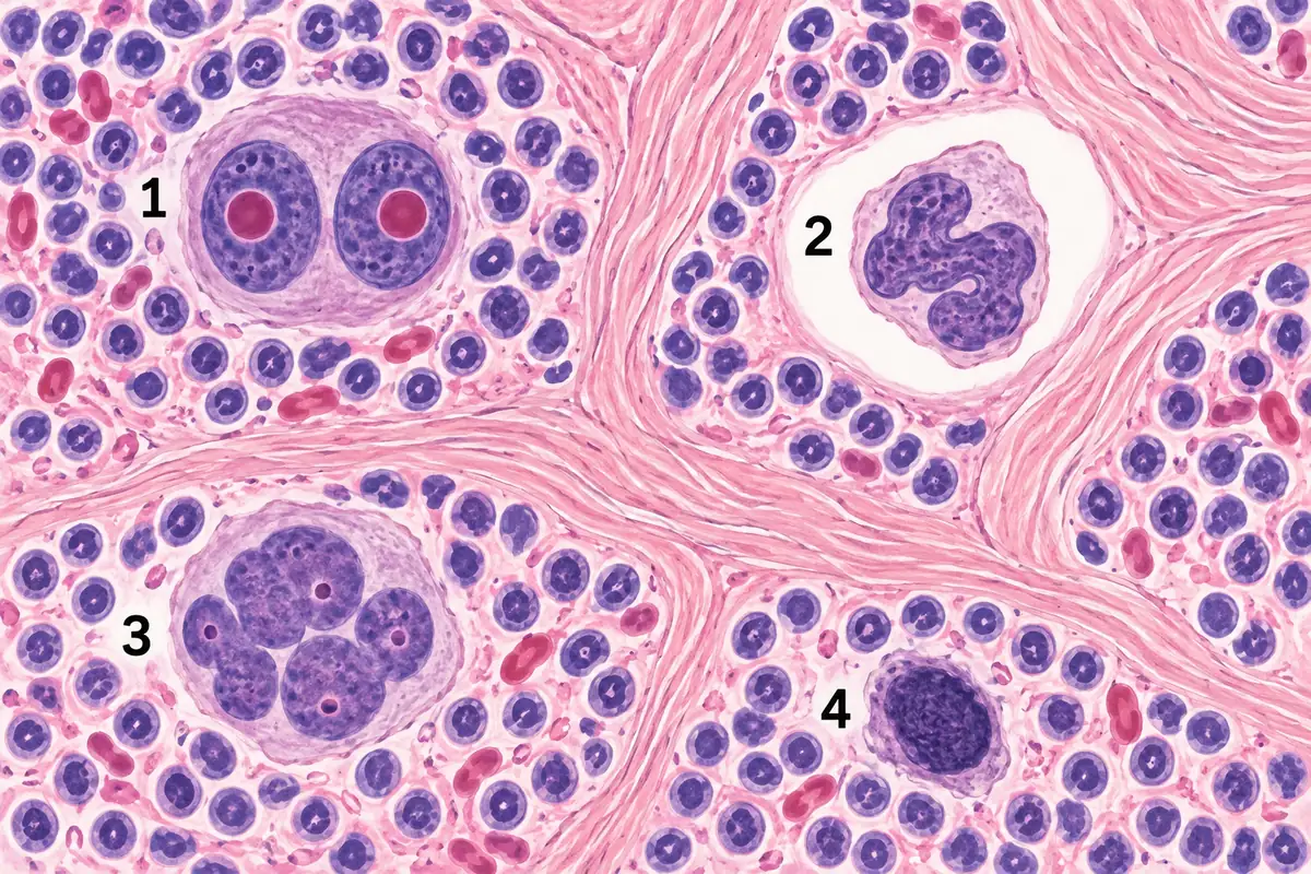
Four large atypical cells from lymph node sections are drawn in the figure. Which is the variant characteristic of the nodular sclerosis type of classical Hodgkin lymphoma?
Look first: look at what surrounds each cell, not only at its nucleus.
 View larger ↗⤢ Tap to enlarge
- ACell 3, the popcorn cell
- BCell 4, the mummified cell
- CCell 2, the lacunar cell
- DCell 1, the lacunar cell
Show the answer and explanation
Answer: C. Cell 2, the lacunar cell
Rule: a cell sitting in a hole is the lacunar cell. Its cytoplasm retracts in formalin-fixed tissue, leaving it in a clear space; cell 2 also has the folded, multilobed nucleus. The collagen bands dividing the node into nodules complete the nodular sclerosis pattern, the commonest type in the West, typically in young women with a mediastinal mass.
Why the closest rival loses: cell 1 (D) is mislabelled in its option: the owl-eye cell with two mirror-image nuclei is the classical Reed-Sternberg cell. It is diagnostic of Hodgkin lymphoma but not of one type. The popcorn cell (A) belongs to nodular lymphocyte-predominant Hodgkin lymphoma, which is not classical, and the mummified cell (B) is a dying tumour cell seen in several types.
Recallab question 55261 · Pathology › Haematology: WBC & Lymphoma · reference: Robbins, Cotran & Kumar Pathologic Basis of Disease
The one-minute routine
| If you see | Look first at | It usually settles |
|---|---|---|
| A Gram film | The colour, then pairs, chains or clusters | The organism, and whether it is inside cells |
| A yeast after serum | Where the filament leaves the cell | Germ tube (Candida albicans) or pseudohypha |
| Stool cysts | The number of nuclei, then fibrils | Entamoeba histolytica, Entamoeba coli or Giardia |
| A red cell film | Inclusions and the background | Asplenia, iron deficiency, megaloblastic or spherocytic |
| A neutrophil | The number of lobes, then the granules | Pelger-Huet, hypersegmentation or toxic change |
| A large cell in a node | What surrounds it | Lacunar, Reed-Sternberg or popcorn cell |
For wounds, weapons and time since death, see forensic medicine spotters for NEET PG. For other kinds of figure, see ten NEET PG image questions. For the eye and the ear, nose and throat, see six eye and ENT spotters. To choose what to practise first, see high impact topics for NEET PG.
Why candidates use Recallab
- Built towards 22,000 — a batch at a time
- Illustrations drawn for the question
- Every option explained, not just the right one
- AI study tools that are live today
- Start free, today
Or see the plans, or try a timed mock section.
Register to practise NEET PG microbiology and pathology questionsFree account, no card needed. Practise with every option explained and a reference on every question.Drawn from questions #55022, #54723, #56995, #55055, #55258, #55261 in the Recallab bank, each with its own verified answer and current-edition reference. Last reviewed 30 September 2026.