
Recallab › Blog › Labour and delivery for NEET PG: six figures from the first stage to the placenta
Labour and delivery for NEET PG: six figures from the first stage to the placenta
A labour figure is solved by placing yourself in the labour first. Work out the stage, find the landmark the drawing is built around, then ask what it calls for now.
Register to practise NEET PG obstetrics questions →Free account, no card needed.
These six illustrated Recallab questions follow one labour from the first stage to the delivery of the placenta: a labour chart, the turn of the head, a vaginal examination, a monitor strip, a vacuum cup and the placenta itself. Whether you are learning obstetrics in MBBS or revising for NEET PG or INI-CET, try each question before opening its worked answer.
Before you read a labour figure: three questions
- Which stage of labour is drawn? The active first stage, full dilatation or the wait for the placenta each asks something different.
- What is the landmark? A time limit on a chart, the pelvic floor, a fontanelle, a stimulus marker or a gap in the membranes.
- Is it from the mother's side? Positions are named from her pelvis, and a chart is read against her own limits, not the examiner's view or habit.
Read the chart, the turn and the fontanelles
Recallab example 1 · a flat line at seven centimetres
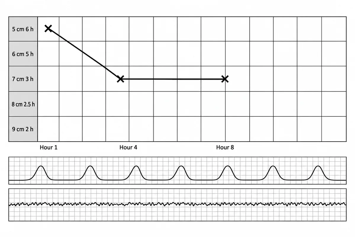
A woman in her first labour at forty weeks is being followed on the labour care guide drawn in the figure, with her vaginal examinations plotted. What does the guide call for now? The cervix section of a labour care guide with three examinations plotted.
Look first: count the hours she has spent at her last dilatation and compare them with the limit printed beside it.
 View larger ↗⤢ Tap to enlarge
- AContinue to observe
- BAssess the cause of delay
- CStart oxytocin augmentation
- DImmediate caesarean section
Show the answer and explanation
Answer: B. Assess the cause of delay
B. Assess the cause of delay. The labour care guide counts the active first stage from five centimetres and gives each centimetre its own time limit, reflecting the slower progress now accepted as normal. She has stayed at seven centimetres from hour four to hour eight, four hours against a limit of three. Passing a limit triggers a review of contractions, position, descent, moulding, hydration and fetal condition, and the plan follows from what that review finds.
Why the other options lose: Continue to observe (A) ignores a limit already passed; the guide has no alert or action line to wait for. Start oxytocin augmentation (C) is one possible plan after the assessment, not an automatic step. Immediate caesarean section (D) is likewise only one possible outcome of the review. From five centimetres the limits run six, five, three, two and a half and two hours.
Recallab question 56179 · Obstetrics › Normal Labour · reference: DC Dutta's Textbook of Obstetrics
Recallab example 2 · where the head turns
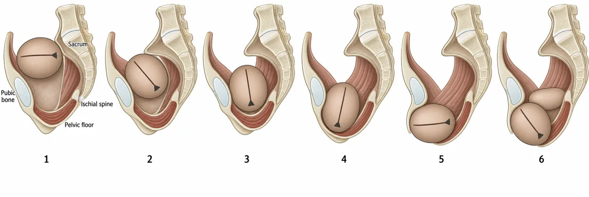
Six moments of a normal vertex delivery are drawn in the figure in the order they happen. At which numbered frame does the back of the head turn forwards to lie behind the pubic bone? The pelvis in section at six moments of one vertex delivery.
Look first: follow the small fontanelle from frame to frame and note the level of the pelvic floor in each.
 View larger ↗⤢ Tap to enlarge
- AFrame 6
- BFrame 2
- CFrame 5
- DFrame 3
Show the answer and explanation
Answer: D. Frame 3
D. Frame 3. At the brim the head lies across the pelvis, because that is the widest diameter there; lower down the widest diameter runs front to back, so the head must turn. It turns where it meets the muscular pelvic floor at the level of the spines: the gutter of the levator muscles slopes down and forwards, and the leading part slides forwards down that slope, carrying the occiput under the pubis. The floor turns the head; the brim only lets it in.
Why the other options lose: Frame 6 (A) is where the shoulders turn and the head follows them further round. Frame 2 (B) shows the head still descending with the occiput to one side, before it has reached the pelvic floor. Frame 5 (C) is the head untwisting after delivery, which follows the turn rather than causing it.
Recallab question 56372 · Obstetrics › Normal Labour & Delivery · reference: DC Dutta's Textbook of Obstetrics
Recallab example 3 · two fontanelles from the foot of the bed
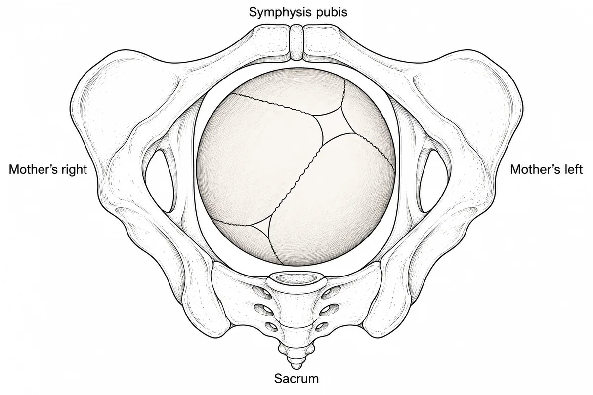
A woman of 30 in her second labour has been fully dilated for an hour, with the head at station 0 and little caput. Her vaginal examination findings are drawn in the figure as the examiner sees them, facing her in the lithotomy position. What is the position of the fetal head? The fetal vertex at full dilatation, drawn from the examiner's side.
Look first: count the sutures meeting at each fontanelle, then name the side from the mother's labels, not your own.
 View larger ↗⤢ Tap to enlarge
- ALeft occipitoanterior
- BLeft occipitoposterior
- CRight occipitoanterior
- DRight occipitoposterior
Show the answer and explanation
Answer: D. Right occipitoposterior
D. Right occipitoposterior. The small triangular fontanelle, where three sutures meet, marks the occiput; the large diamond, where four meet, is the front of the head. Here the triangle lies towards the sacrum, so the occiput is posterior, and facing her, the examiner's left-hand side is the mother's right. The occiput therefore sits in her right posterior quadrant. Both fontanelles can be felt because an occipitoposterior head is usually deflexed.
Why the other options lose: Left occipitoanterior (A) takes the diamond for the occiput; the diamond is the sinciput, lying anteriorly on her left. Left occipitoposterior (B) finds the right fontanelle but names the side from the examiner's view instead of the mother's. Right occipitoanterior (C) makes both errors at once. Three sutures, triangle, occiput; four sutures, diamond, front of the head.
Recallab question 69842 · Obstetrics › Malpresentation · reference: DC Dutta's Textbook of Obstetrics
Read the strip, the cup and the placenta
Recallab example 4 · a flat strip and a stimulus
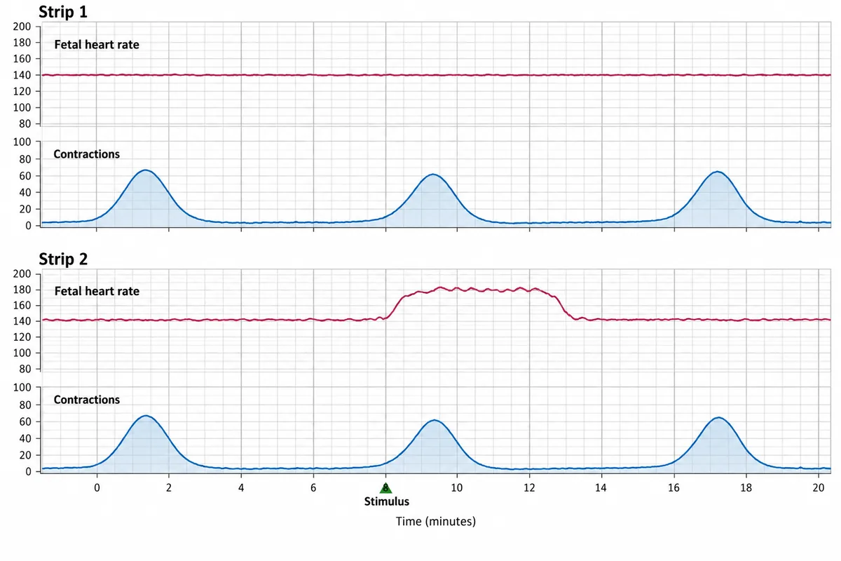
Two strips from the same labour are drawn in the figure, the second recorded immediately after a firm stimulus was applied at a vaginal examination. What does the change between the strips mean? Two monitor strips from one labour, the second recorded after a stimulus.
Look first: compare the baseline of strip 1 with what happens in strip 2 just after the marker.
 View larger ↗⤢ Tap to enlarge
- AScalp pH likely normal
- BFetus merely asleep
- CFetus acidotic
- DTest invalid before second stage
Show the answer and explanation
Answer: A. Scalp pH likely normal
A. Scalp pH likely normal. Strip 1 has a flat baseline with reduced variability and no accelerations. Strip 2 shows a clear rise beginning immediately after the stimulus marker, and an acceleration of that size and length provoked by stimulating the presenting part predicts a normal scalp pH, so fetal blood sampling can be avoided. The reverse inference does not hold: a trace that stays flat after stimulation still needs sampling or delivery.
Why the other options lose: Fetus merely asleep (B) would explain the flat first strip, but it does not explain away the reassurance the provoked rise gives. Fetus acidotic (C) is wrong because an acidotic fetus does not mount an acceleration to stimulation, which is exactly why the test works. Test invalid before second stage (D) is wrong because it is used through labour once the cervix admits an examining finger. A rise on stimulation reassures; no rise does not condemn.
Recallab question 58275 · Obstetrics › Abnormal Labour & Obstetric Emergencies · reference: Williams Obstetrics
Recallab example 5 · four crosses on the vertex
A primigravida of 26 has been fully dilated and pushing for two hours. The head is at +2 station in a direct occipitoanterior position with little caput, and the fetal heart shows prolonged decelerations. A vacuum birth is chosen. Four positions for the centre of the cup on the fetal vertex are drawn in the figure. Where should the cup be applied? The top of the fetal head with four numbered crosses.
Look first: check whether each cross lies on the sagittal suture, then how far it sits from each fontanelle.
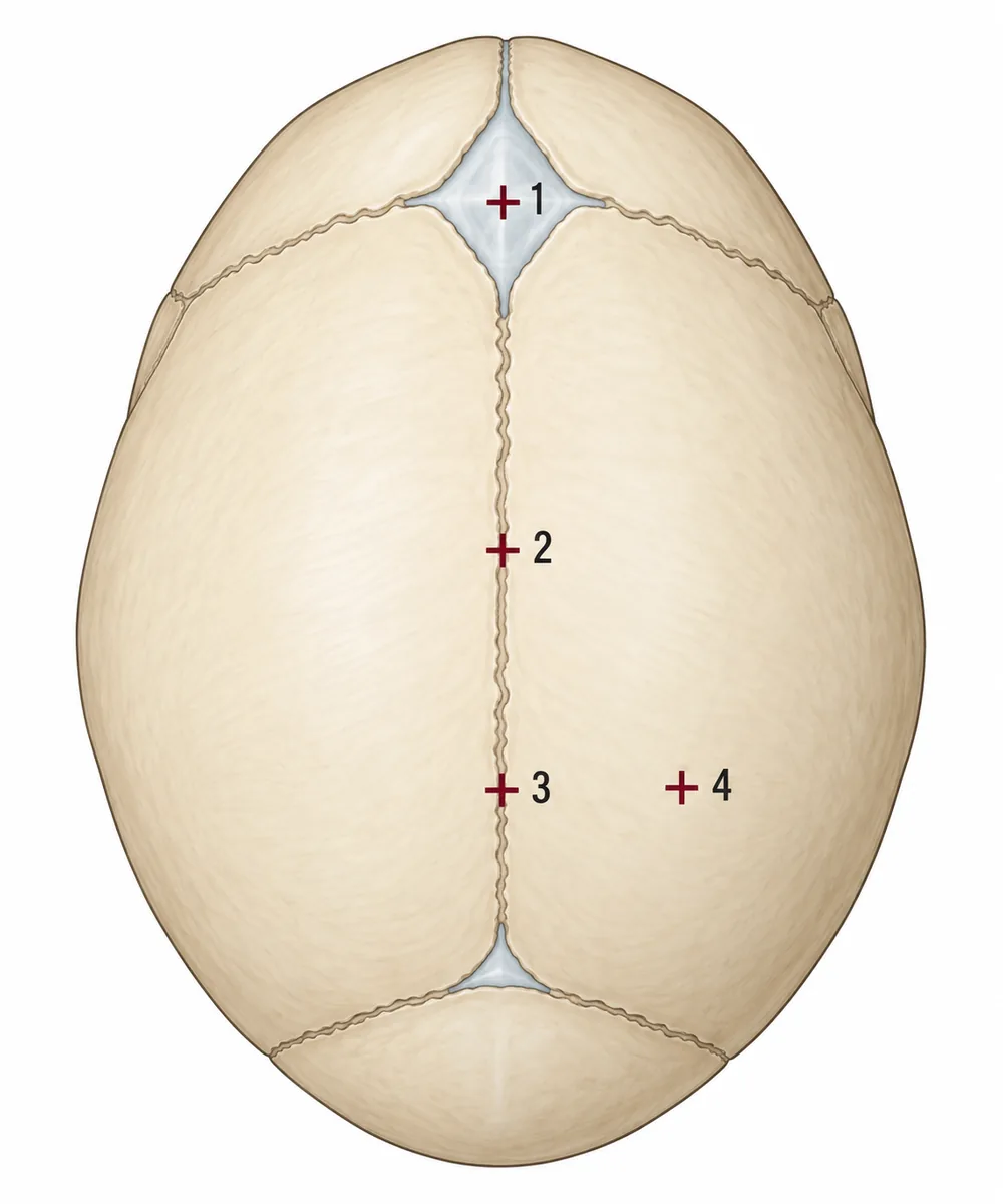 View larger ↗⤢ Tap to enlarge
- APosition 3
- BPosition 4
- CPosition 2
- DPosition 1
Show the answer and explanation
Answer: A. Position 3
A. Position 3. The cup is centred on the flexion point: on the sagittal suture about 3 cm in front of the posterior fontanelle. The fontanelles are about 9 cm apart, so that is a third of the way forward, and a standard 6 cm cup placed there has its rim just at the posterior fontanelle. Traction through the flexion point flexes the head so that it presents its smallest diameter, the suboccipitobregmatic, and correct placement lowers the risk of the cup detaching and of scalp injury.
Why the other options lose: Position 4 (B) is off the midline, so traction tilts the head and causes asynclitism. Position 2 (C) is on the suture but too far forward, so traction still deflexes the head slightly. Position 1 (D) sits on the anterior fontanelle; traction there deflexes the head and presents a wider diameter. Cup rim on the posterior fontanelle, centre on the suture.
Recallab question 69251 · Obstetrics › Operative Obstetrics · reference: DC Dutta's Textbook of Obstetrics
Recallab example 6 · the membranes as well as the disc
A delivered placenta is drawn in the figure from both surfaces with the membranes spread out. Which finding indicates that something has been retained? One delivered placenta drawn from both surfaces with the membranes spread out.
Look first: check the membranes as well as the disc: follow every vessel to where it ends.
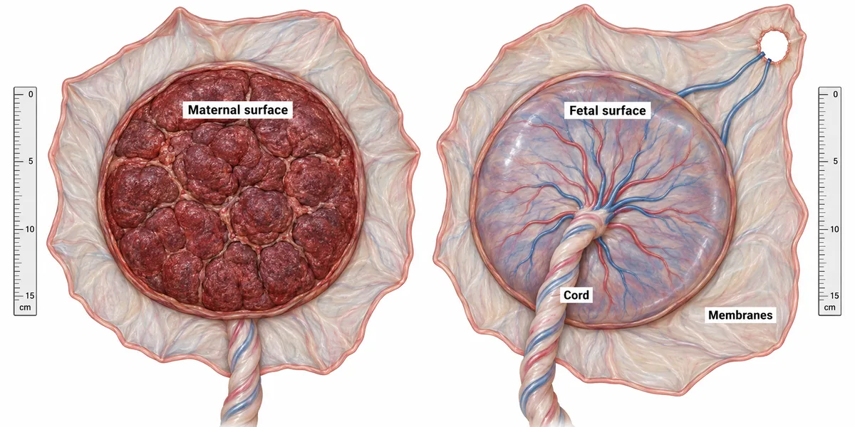 View larger ↗⤢ Tap to enlarge
- AA cord inserted off centre
- BA complete maternal surface
- CCalcification of the maternal surface
- DVessels ending at a hole in the membranes
Show the answer and explanation
Answer: D. Vessels ending at a hole in the membranes
D. Vessels ending at a hole in the membranes. Vessels that run off the edge of the disc and stop at a hole in the membranes mean a separate lobe has been torn off and left inside. Such an accessory lobe sits apart in the membranes, joined only by its vessels, so the cut vessel ends at the margin of the defect are the evidence. Retained tissue stops the uterus contracting down, so bleeding continues, and it is a focus for infection days later, with offensive lochia and fever.
Why the other options lose: A cord inserted off centre (A) is a normal variant. A complete maternal surface (B) can still be missing an accessory lobe: the disc being whole is exactly what makes this trap worth knowing. Calcification of the maternal surface (C) is a normal finding at term and retains nothing. Check the membranes as well as the disc.
Recallab question 55094 · Obstetrics › Normal labour and delivery · reference: DC Dutta's Textbook of Obstetrics
The one-minute routine
| If the figure gives | Ask first | It settles |
|---|---|---|
| A labour care guide with examinations plotted | How long at this centimetre, against its own limit? | A limit passed means assess the cause and plan |
| The head drawn at stages of its descent | Where does it meet the sloping pelvic floor? | Internal rotation brings the occiput under the pubis there |
| Fontanelles felt at full dilatation | Where is the three-suture triangle, on the mother's side? | The position of the occiput |
| A flat strip, then a stimulus | Does the heart rate rise after the marker? | A provoked rise predicts a normal scalp pH |
| Crosses on the vertex for a vacuum cup | On the suture, about 3 cm in front of the posterior fontanelle? | The flexion point |
| A delivered placenta | Do any vessels end at a hole in the membranes? | A retained accessory lobe, even with a complete disc |
Practise obstetrics questions, including normal labour and malpresentation, and see six pictures from earlier in pregnancy in antenatal care figures.
Why candidates use Recallab
- Built towards 22,000 — a batch at a time
- Illustrations drawn for the question
- Every option explained, not just the right one
- AI study tools that are live today
- Start free, today
Or see the plans, or try a timed mock section.
Register to practise NEET PG obstetrics questionsFree account, no card needed. Practise with every option explained and a reference on every question.Drawn from questions #56179, #56372, #69842, #58275, #69251, #55094 in the Recallab bank, each with its own verified answer and current-edition reference. Last reviewed 21 October 2026.