
Recallab › Blog › Pathology for NEET PG: six figures on tumour invasion, grade and stage
Pathology for NEET PG: six figures on tumour invasion, grade and stage
A tumour figure asks more than whether the cells look abnormal. Read the architecture, trace the boundary, then separate grade from stage.
Register to practise NEET PG pathology questions →Free account, no card needed.
These six illustrated Recallab questions take you from a benign mimic to invasion, routes of spread and the report that follows. Whether you are learning pathology in MBBS or revising for NEET PG or INI-CET, try each question before opening its worked answer.
Before you call it invasive: three questions
- What is the architecture? Read gland arrangement, pigment and location alongside the nuclei.
- Where is the boundary? Trace the basement membrane or outer cell layer, then look for cells beyond it.
- Is the question about appearance or extent? Histological scores decide grade; spread into surrounding structures changes stage.
Benign mimics and the boundary of invasion
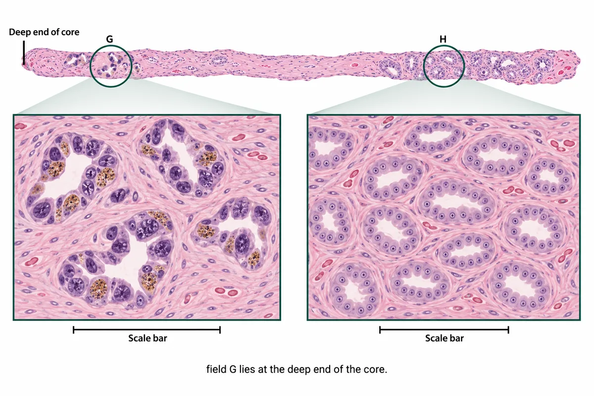
Recallab example 1 · large nuclei at the deep end
A biopsy core is drawn in the figure with two groups of glands enlarged at one magnification. One group has strikingly large dark nuclei. What are those glands? One biopsy core with two groups of glands enlarged at one magnification, one group carrying strikingly large dark nuclei.
Look first: compare the gland arrangement, cytoplasmic pigment and position in the core before judging nuclear size.
 View larger ↗⤢ Tap to enlarge
- AForeign body reaction
- BNormal structure at the deep end
- CEarlier treatment effect
- DHighest-grade tumour in the core
Show the answer and explanation
Answer: B. Normal structure at the deep end
B. Normal structure at the deep end. Field G represents seminal vesicle or ejaculatory duct tissue sampled at the deep end of a prostate core. Its coarse golden brown pigment, scattered glands and widely varying dark nuclei explain the alarming appearance. Field H shows the contrasting tumour pattern: small crowded glands with more uniform, pale nuclei. Nuclear size alone would send you towards the wrong group.
Why the other options lose: Foreign body reaction (A) would involve a reaction around the material; here the pigment is inside the gland lining cells and the surrounding tissue shows no reaction. Earlier treatment effect (C) produces gland shrinkage and clearer cytoplasm, rather than this golden pigment. Highest-grade tumour in the core (D) mistakes normal tissue for the most aggressive tumour: position, pigment and gland arrangement identify the benign mimic.
Recallab question 59719 · Pathology › Male Genital Tract · reference: Robbins, Cotran & Kumar Pathologic Basis of Disease
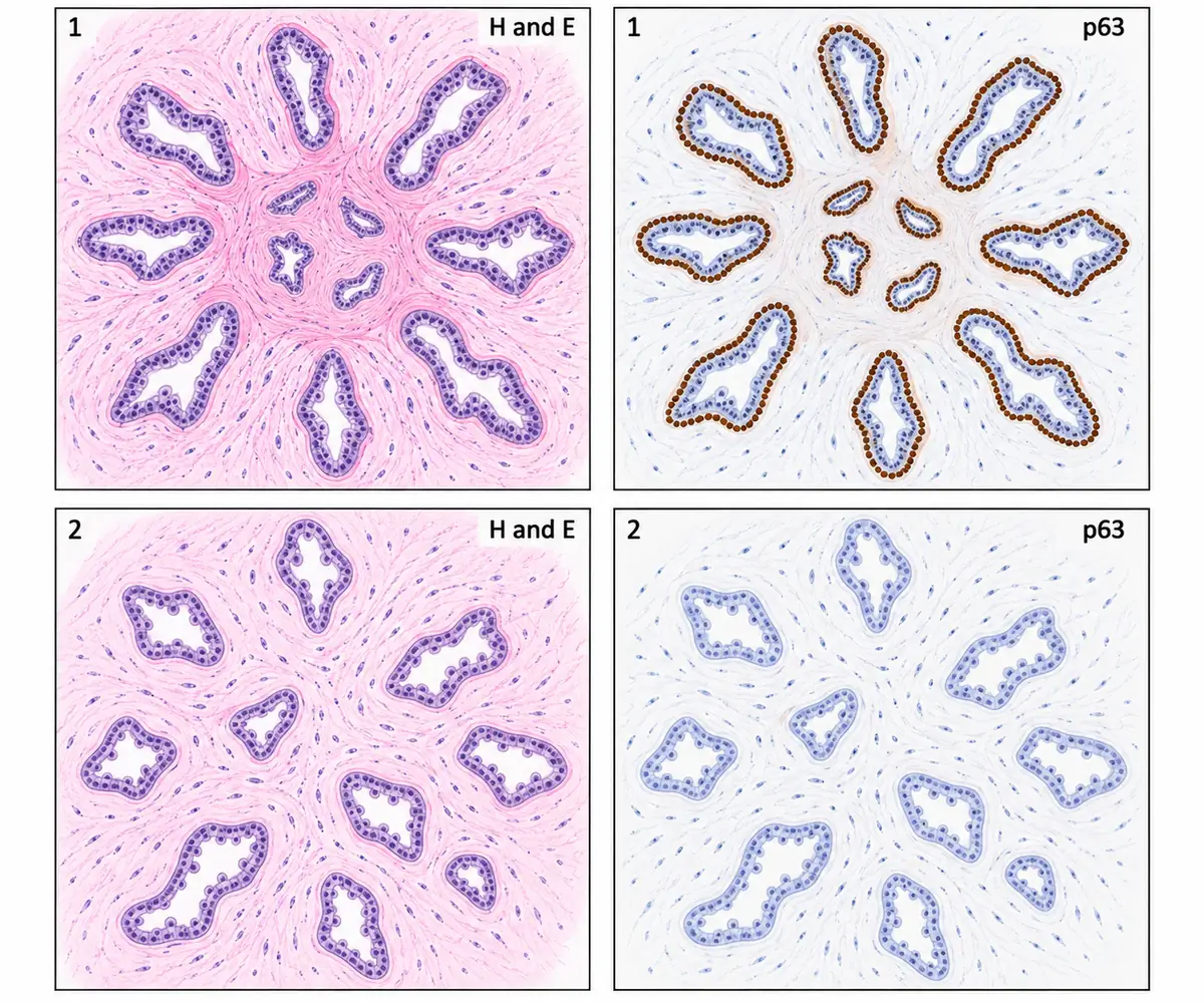
Recallab example 2 · two star-shaped breast lesions
Screening mammography in a woman of 52 shows a small stellate density, and a core biopsy is taken. Two stellate breast lesions are drawn in the figure, each in routine stain and in a p63 immunostain. Which finding proves that one of the two lesions is benign? Two stellate breast lesions, each in routine stain and in a p63 immunostain, at the same magnification.
Look first: follow the p63-stained nuclei around each gland, including the distorted glands in the centre.
 View larger ↗⤢ Tap to enlarge
- AOpen lumina with apical snouts
- BIntact myoepithelial cell layer
- CA central core without glands
- DAngulated glands trapped in scar
Show the answer and explanation
Answer: B. Intact myoepithelial cell layer
B. Intact myoepithelial cell layer. In lesion 1, p63 stains a continuous outer ring of myoepithelial nuclei around every gland, including those trapped in the central scar. With the scarred architecture shown, that retained layer supports a radial scar. Lesion 2 has lost the layer and shows the open, angulated glands of tubular carcinoma. Both are stellate, so the overall shape does not settle the question.
Why the other options lose: Open lumina with apical snouts (A) describe the tubular carcinoma in lesion 2, whose glands lack the p63 ring. A central core without glands (C) misses the trapped glands within the radial scar; the decisive finding is their retained myoepithelial layer. Angulated glands trapped in scar (D) can mimic invasion and cannot establish benignity by shape alone.
Recallab question 57922 · Pathology › Breast · reference: Robbins, Cotran & Kumar Pathologic Basis of Disease
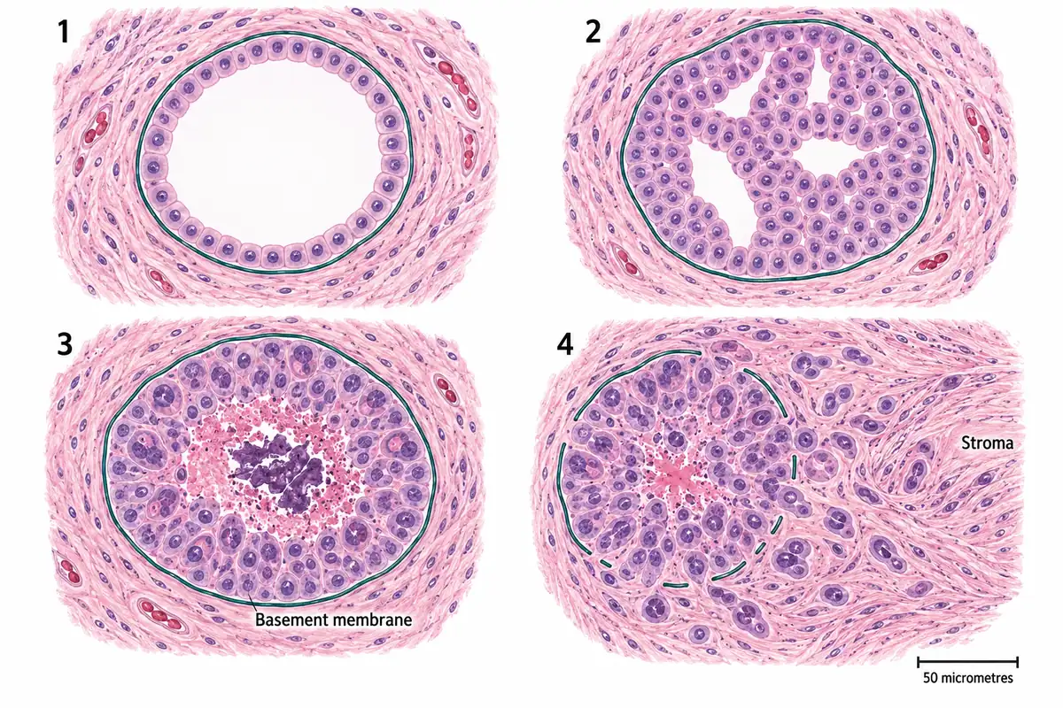
Recallab example 3 · trace the basement membrane
Four breast lesions are drawn in the figure at one magnification with the basement membrane traced in each. Which lesion has become invasive? Four breast lesions at one magnification with the basement membrane traced.
Look first: trace the boundary of each duct and check whether cells lie beyond it in the stroma.
 View larger ↗⤢ Tap to enlarge
- ALesion 4
- BLesion 2
- CLesion 1
- DLesion 3
Show the answer and explanation
Answer: A. Lesion 4
A. Lesion 4. Its basement membrane is broken and tumour cells extend into the surrounding stroma as cords and single cells. That crossing defines invasion. Lesion 3 has equally malignant-looking cells and central necrosis, but its intact basement membrane keeps the process in situ. Judge the boundary before judging how alarming the nuclei look.
Why the other options lose: Lesion 2 (B) shows hyperplasia: regular cells fill and expand a duct while its membrane remains intact. Lesion 1 (C) is a normal duct, with one regular cell layer inside an intact membrane. Lesion 3 (D) is carcinoma in situ: pleomorphism and necrosis do not establish invasion while the cells remain confined behind the membrane.
Recallab question 55157 · Pathology › Reproductive & Breast Pathology · reference: Robbins, Cotran & Kumar Pathologic Basis of Disease
Routes of spread, grade and stage
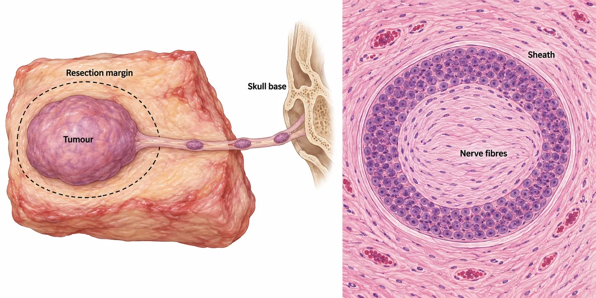
Recallab example 4 · a route towards the skull base
A carcinoma of the cheek was excised and every margin was reported clear of tumour, yet within the year it returned along a line running backwards towards the base of the skull. The map of the specimen and one field taken from it are drawn in the figure. What does the field show? A map of the resected specimen with the line of later recurrence marked, and one microscopic field taken from the edge of the resection.
Look first: locate the tumour cells relative to the sheath and fibres in the enlarged field, then follow that structure on the specimen map.
 View larger ↗⤢ Tap to enlarge
- APerineural tumour spread
- BTumour displaced by handling
- CLymphatic tumour emboli
- DArterial tumour invasion
Show the answer and explanation
Answer: A. Perineural tumour spread
A. Perineural tumour spread. The field shows tumour cells encircling nerve fibres inside the outer sheath. This plane offers a route away from the main mass. Separate deposits can lie along the nerve with normal segments between them, beyond the tissue removed. The line of recurrence towards the skull base therefore fits spread along the nerve despite the reported clear margins.
Why the other options lose: Tumour displaced by handling (B) does not explain the organised position within the nerve sheath and recurrence along its course. Lymphatic tumour emboli (C) would lie in lymphatic channels and favour spread to draining nodes, rather than this nerve-shaped route. Arterial tumour invasion (D) would place cells in an arterial lumen and provide a route to distant sites; the field instead shows cells around nerve fibres within their sheath.
Recallab question 58223 · Pathology › Neoplasia · reference: Robbins, Cotran & Kumar Pathologic Basis of Disease
Recallab example 5 · add the three histological scores
Three fields from one invasive ductal carcinoma of the breast are drawn in the figure, each assessed for one feature and given a score. What is the tumour's histological grade? Three fields from one breast carcinoma, each with the feature assessed and its score.
Look first: read the score under each field and add all three before choosing the grade.
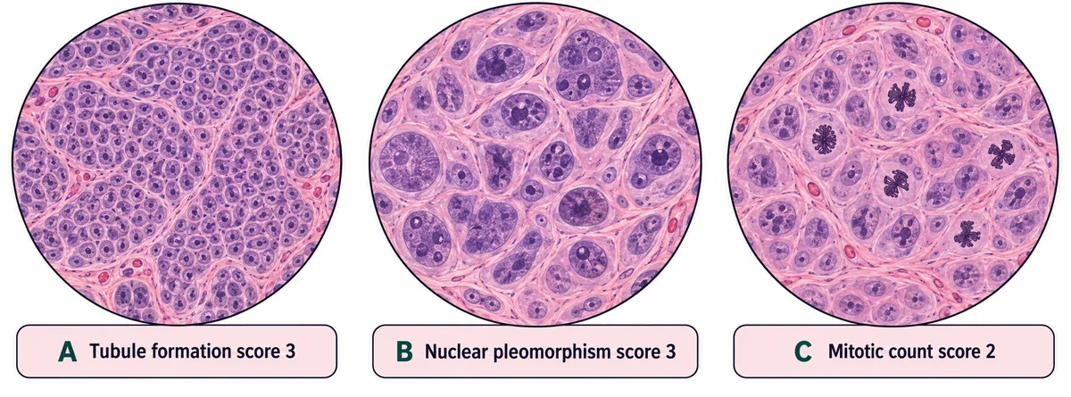 View larger ↗⤢ Tap to enlarge
- AGrade 3, highest score 3
- BGrade 2, total score 8
- CNot gradable without staging
- DGrade 3, total score 8
Show the answer and explanation
Answer: D. Grade 3, total score 8
D. Grade 3, total score 8. Nottingham grading adds the scores for tubule formation, nuclear pleomorphism and mitotic count. Here the total is 3 + 3 + 2 = 8. Totals of 3–5 give grade 1, 6–7 give grade 2, and 8–9 give grade 3. Grade describes the histological appearance; it can be assigned independently of stage.
Why the other options lose: Grade 3, highest score 3 (A) reaches the right grade for the wrong reason: the sum of all three features determines it, rather than the highest individual score. Grade 2, total score 8 (B) adds correctly but uses the wrong range; grade 2 covers totals of 6 or 7. Not gradable without staging (C) confuses histological appearance with extent: size and nodal involvement inform stage, but are unnecessary for this grade calculation.
Recallab question 56117 · Pathology › Breast · reference: Robbins, Cotran & Kumar Pathologic Basis of Disease
Recallab example 6 · same tumour, different extent
Two kidneys were removed from two patients for a tumour of the same kind, and both are drawn in the figure in section with a microscopic view of each beside it and a strip beneath showing how the categories are assigned. Which line of the report differs between the two? Two kidney specimens in section holding tumours of the same size and appearance, one with a solid growth filling the vein at the hilum, with the strip of categories printed beneath.
Look first: compare size and microscopic appearance first, then check the vein leaving each kidney.
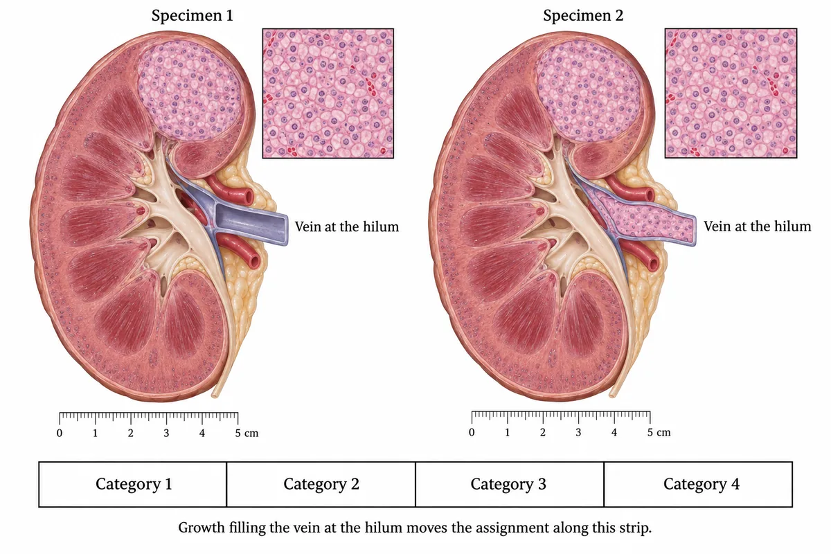 View larger ↗⤢ Tap to enlarge
- AThe tumour size
- BThe stage category
- CThe diagnosis
- DThe grade
Show the answer and explanation
Answer: B. The stage category
B. The stage category. The tumours have the same diameter and identical microscopic appearances. The difference is the solid extension of tumour into the renal vein in specimen 2. That changes the extent of disease and therefore the stage category. It does not change the tumour identity or its histological grade.
Why the other options lose: The tumour size (A) is identical in the two drawings. The diagnosis (C) remains the same because extending into a vein does not turn the tumour into a different type. The grade (D) describes microscopic appearance, which the matching insets deliberately hold constant. Ask separately what the tumour is, how it looks and how far it has spread.
Recallab question 59417 · Pathology › Kidney · reference: Robbins, Cotran & Kumar Pathologic Basis of Disease
The one-minute routine
| If the figure gives | Ask first | It settles |
|---|---|---|
| Large dark nuclei in a prostate core | Where are the glands, and is there golden pigment? | A normal deep structure can mimic tumour |
| Two stellate breast lesions | Does p63 ring the glands? | A retained myoepithelial layer supports the radial scar shown |
| Malignant-looking cells within a duct | Have cells crossed the basement membrane? | In situ disease or invasion |
| Recurrence along a line towards the skull base | Are tumour cells inside a nerve sheath? | Perineural spread |
| Three breast tumour scores | What is the sum of tubules, nuclei and mitoses? | 3–5: grade 1; 6–7: grade 2; 8–9: grade 3 |
| Equal-sized kidney tumours with matching histology | Does tumour extend into the renal vein? | A different stage category despite the same diagnosis and grade |
Practise pathology questions and compare other illustrations in ten NEET PG image questions.
Why candidates use Recallab
- Built towards 22,000 — a batch at a time
- Illustrations drawn for the question
- Every option explained, not just the right one
- AI study tools that are live today
- Start free, today
Or see the plans, or try a timed mock section.
Register to practise NEET PG pathology questionsFree account, no card needed. Practise with every option explained and a reference on every question.Drawn from questions #59719, #57922, #55157, #58223, #56117, #59417 in the Recallab bank, each with its own verified answer and current-edition reference. Last reviewed 14 October 2026.