
Recallab › Blog › Surgery for NEET PG: six figures that decide the gastrointestinal operation
Surgery for NEET PG: six figures that decide the gastrointestinal operation
The diagnosis starts the decision. The anatomy in the figure tells you what to remove, what to drain and what may be worth preserving.
Register to practise NEET PG surgery questions →Free account, no card needed.
These six Recallab MentorQ questions use teaching illustrations to connect a clinical problem to its treatment. Whether you are learning abdominal anatomy, working through an internship or revising after graduation, start by naming the structure and describing what has changed. Try each question before opening the worked answer.
Before you choose an operation: three questions
- Where is the problem? Locate the abnormal bowel loop, the depth of invasion or the dilated part of a duct.
- What tissue remains usable? Check bowel viability, excision margins and how much duct wall is intact.
- What must treatment achieve? Remove a segment at risk, relieve obstruction, reconstruct drainage or preserve tissue for reassessment.
Bowel: position, viability and invasion depth
Recallab example 1 · a bowel loop out of place
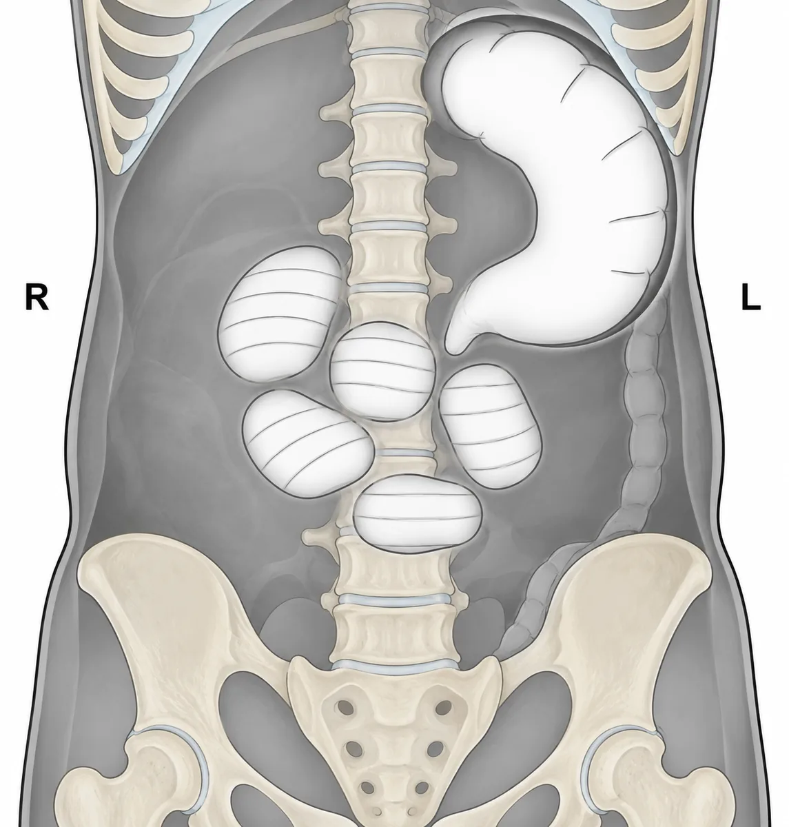
A woman of 38 has had 30 hours of colicky abdominal pain, distension and vomiting, and has passed no flatus since last night. She had a caesarean section four years ago. She is not peritonitic and her lactate is normal. Her supine abdominal film is drawn in the figure. What is the preferred definitive treatment? Her supine abdominal film, drawn as a line diagram with the patient's right and left marked.
Look first: locate the largest loop, follow its taper and check which parts of the bowel are empty.
 View larger ↗⤢ Tap to enlarge
- ASigmoidoscopic decompression
- BRight hemicolectomy
- CColonoscopic detorsion
- DAdhesiolysis alone
Show the answer and explanation
Answer: B. Right hemicolectomy
B. Right hemicolectomy. The large loop in the left upper abdomen tapers towards an empty right iliac fossa. Together with dilated small bowel and a collapsed distal colon, this identifies a caecal volvulus: a mobile caecum has twisted and moved across the abdomen. Right hemicolectomy removes the mobile segment and provides definitive treatment with the least recurrence. A normal lactate and the absence of peritonitis do not make endoscopic untwisting the preferred treatment.
Why the other options lose: Sigmoidoscopic decompression (A) treats a sigmoid volvulus, whose loop rises from the pelvis; the sigmoid here is empty. Colonoscopic detorsion (C) rarely succeeds for a caecal volvulus and can delay the operation. Adhesiolysis alone (D) would address an adhesive band, but the film shows a displaced, twisted caecum. Her earlier abdominal operation is a tempting clue; the anatomy on the film decides the answer.
Recallab question 69001 · Surgery › Intestinal Obstruction · reference: Bailey & Love's Short Practice of Surgery
Recallab example 2 · bowel that may recover
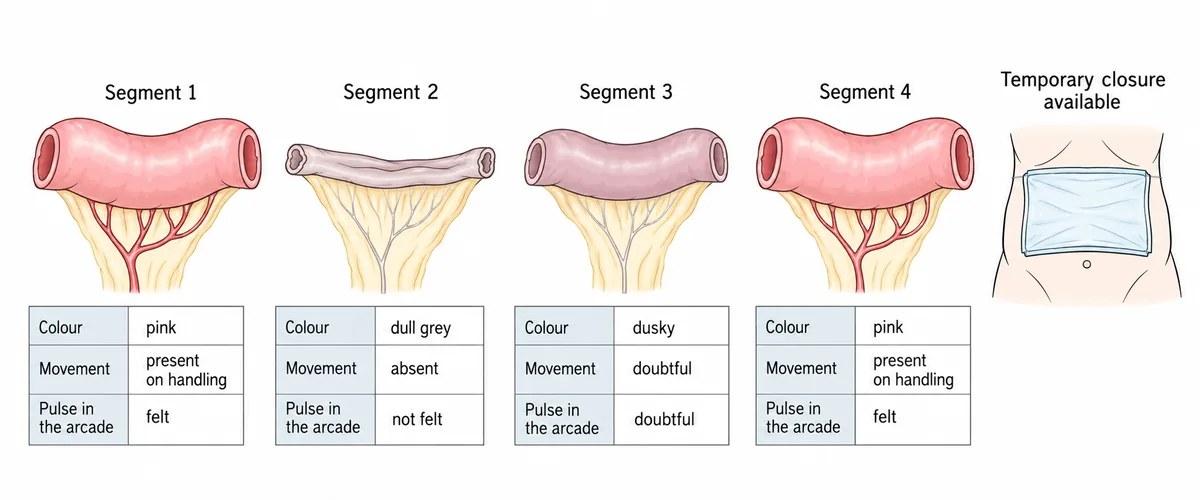
At operation for a blocked mesenteric vessel the blood flow has been restored and the bowel has been watched for ten minutes. The figure records the colour, the movement and the pulse in the arcade for four segments. What should be done with segment 3? Four segments of small bowel after flow was restored, with three observations recorded for each.
Look first: read all three observations for segment 3 and compare them with the clearly healthy and clearly unhealthy segments.
 View larger ↗⤢ Tap to enlarge
- APlanned second-look laparotomy
- BPrimary anastomosis across it
- CExteriorise it as a stoma
- DResect it now
Show the answer and explanation
Answer: A. Planned second-look laparotomy
A. Planned second-look laparotomy. Segment 3 is dusky, with doubtful movement and a doubtful arcade pulse. Its viability remains uncertain after only ten minutes of restored blood flow. Leave this segment in place, use temporary abdominal closure and arrange a planned return to assess it again. Bowel that looks doubtful can recover as circulation improves. Segment 2, which fails all three observations, is clearly non-viable and needs removal; segments 1 and 4 look viable.
Why the other options lose: Primary anastomosis across it (B) places a join in tissue whose blood supply is uncertain, risking breakdown. Exteriorise it as a stoma (C) commits the doubtful segment to the skin without resolving whether it can recover. Resect it now (D) removes potentially recoverable bowel and risks unnecessary loss of length. The second look must be planned, rather than postponed until deterioration forces a return.
Recallab question 58963 · Surgery › Small & Large Intestine · reference: Bailey & Love's Short Practice of Surgery
Recallab example 3 · cancer below the stalk
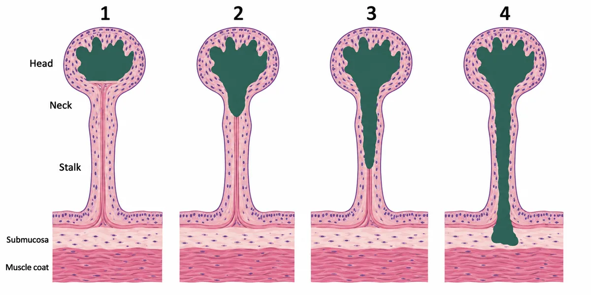
Four stalked colonic polyps, each found to contain an invasive adenocarcinoma, were removed whole by snare. They are drawn in the figure as they sat on the bowel wall, with the cancer shaded. In each, the cancer is well differentiated with no lymphovascular invasion. Which patient needs a segmental colectomy? Four stalked polyps drawn on the bowel wall in section, the cancer shaded in each.
Look first: trace the shaded cancer from the head downwards and identify whether it enters the bowel wall beneath the stalk.
 View larger ↗⤢ Tap to enlarge
- APolyp 3
- BPolyp 1
- CPolyp 2
- DPolyp 4
Show the answer and explanation
Answer: D. Polyp 4
D. Polyp 4. The shaded cancer extends below the stalk into the submucosa of the bowel wall. This is Haggitt level 4 and carries a risk of lymph node spread, so treatment is segmental colectomy with the regional lymph nodes. The favourable differentiation and absence of lymphovascular invasion do not remove the significance of this depth. For levels 1 to 3, favourable histology and a clear excision margin can allow snare removal alone to suffice.
Why the other options lose: Polyp 3 (A) reaches the stalk but stops above the bowel wall: Haggitt level 3. Polyp 1 (B) is confined to the head: level 1. Polyp 2 (C) reaches the neck: level 2. With clear margins and the favourable features given, these three do not need colectomy on invasion depth alone. Do not confuse invasion of the stalk with invasion of the bowel wall beneath it.
Recallab question 56162 · Surgery › Small & Large Intestine · reference: Bailey & Love's Short Practice of Surgery
Pancreas and bile ducts: drainage or reconstruction
Recallab example 4 · a wide pancreatic duct
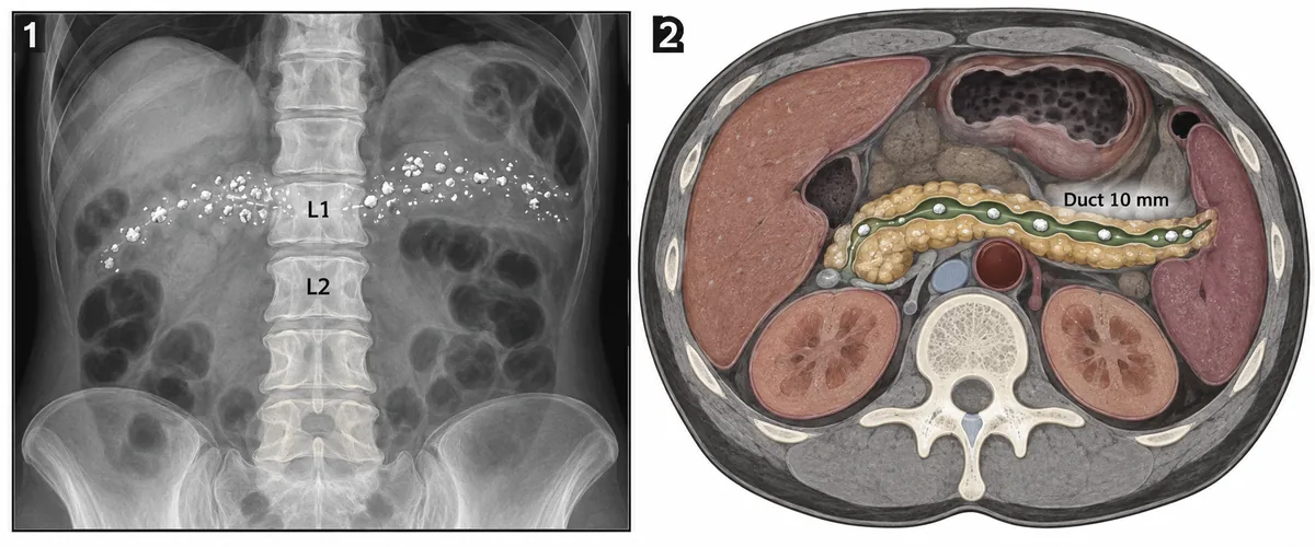
A 42-year-old man who drank heavily for fifteen years has years of epigastric pain boring through to the back, pale bulky stools and new diabetes. The pain persists despite abstinence, enzyme supplements and analgesia. His plain film and CT are drawn in the figure. Which operation suits him? A plain abdominal film and a CT section from one patient, numbered 1 and 2.
Look first: read the duct diameter, follow the distribution of stones and strictures, and check the size of the pancreatic head.
 View larger ↗⤢ Tap to enlarge
- ADistal pancreatectomy
- BPancreaticoduodenectomy
- CLongitudinal pancreaticojejunostomy
- DEndoscopic sphincterotomy
Show the answer and explanation
Answer: C. Longitudinal pancreaticojejunostomy
C. Longitudinal pancreaticojejunostomy. Calcification, pain radiating to the back, steatorrhoea and diabetes fit chronic calcific pancreatitis with exocrine and endocrine failure. The CT shows a 10 mm main duct containing stones and strictures along its length, while the head is not enlarged. Opening the duct longitudinally and joining it side to side to a Roux loop of jejunum drains the obstructed duct and relieves pressure. The wide duct makes drainage suitable in this patient.
Why the other options lose: Distal pancreatectomy (A) removes the tail without draining the rest of the affected duct and may worsen diabetes. Pancreaticoduodenectomy (B) removes the head, but there is no enlarged head or mass to justify that choice here. Endoscopic sphincterotomy (D) opens the outlet at the papilla; it does not address the strictures and stones distributed along the duct. The duct diameter and head size decide the operation.
Recallab question 55759 · Surgery › Hepatobiliary & Pancreas · reference: Bailey & Love's Short Practice of Surgery
Recallab example 5 · a cyst beside the papilla
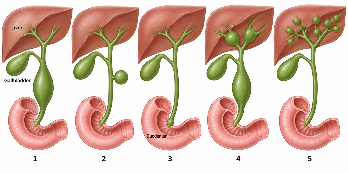
Five biliary trees, each with a different form of choledochal cyst, are drawn in the figure. Which one is usually treated endoscopically through the duodenum rather than by excision? Five biliary trees, each with a different cystic dilatation, numbered 1 to 5.
Look first: locate each dilatation in relation to the liver, the common bile duct and the duodenal wall.
 View larger ↗⤢ Tap to enlarge
- ATree 1
- BTree 2
- CTree 3
- DTree 5
Show the answer and explanation
Answer: C. Tree 3
C. Tree 3. The lowest part of the bile duct bulges into the duodenum at the papilla. This is a choledochocele, Todani type III. Its position within the duodenal wall allows endoscopic sphincterotomy or unroofing through the duodenum. Its cancer risk is lower than that of the extrahepatic cyst types generally treated by excision. First locate the cyst; its relationship to the duodenal lumen explains the treatment route.
Why the other options lose: Tree 1 (A) is a type I dilatation of the common bile duct, requiring excision and biliary reconstruction because drainage alone leaves the cyst lining in place. Tree 2 (B) is a type II diverticulum, which is excised surgically. Tree 5 (D) shows intrahepatic duct dilatations of Caroli disease; treatment may require liver resection when localised or transplantation when diffuse. None has the intraduodenal position shown in tree 3.
Recallab question 56963 · Surgery › The Gall Bladder & Bile Ducts · reference: Bailey & Love's Short Practice of Surgery
Recallab example 6 · how much duct wall remains
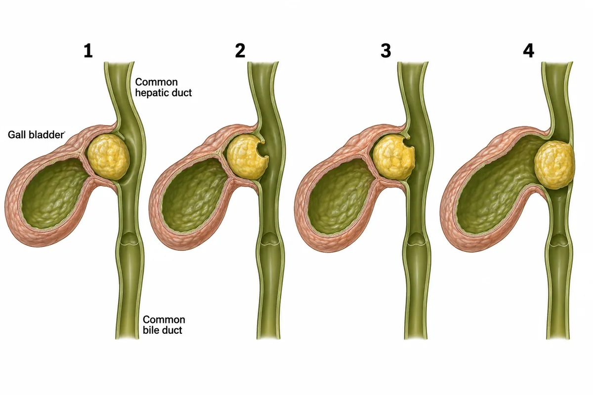
A woman of 80 with a shrunken gall bladder full of stones becomes jaundiced. Four possible findings at the neck of her gall bladder are drawn in the figure. Which finding commits the surgeon to joining the bile duct to the bowel? Four findings at the neck of the gall bladder, drawn at one scale and numbered 1 to 4.
Look first: compare the circumference of intact bile duct wall in all four panels before choosing a repair.
 View larger ↗⤢ Tap to enlarge
- APanel 1
- BPanel 3
- CPanel 4
- DPanel 2
Show the answer and explanation
Answer: C. Panel 4
C. Panel 4. The stone has eroded through the whole circumference of the bile duct, leaving no wall to repair at that level. This is the complete duct destruction of Csendes type IV Mirizzi syndrome. A Roux-en-Y hepaticojejunostomy joins the remaining duct to jejunum to restore bile drainage. The decision follows the amount of duct wall lost, rather than the presence of jaundice alone.
Why the other options lose: Panel 1 (A) shows external compression with an intact duct; removing the gallbladder relieves the obstruction. Panel 3 (B) retains about half the circumference, so reconstruction using a flap of gallbladder wall can be possible in the pattern shown. Panel 2 (D) has a small fistula, which can be closed or patched, including repair over a T-tube. Complete circumferential destruction is the finding that commits this question to a bile duct-to-bowel join.
Recallab question 56764 · Surgery › The Gall Bladder & Bile Ducts · reference: Bailey & Love's Short Practice of Surgery
The one-minute routine
| If the figure gives | Ask first | It settles |
|---|---|---|
| A large bowel loop in the left upper abdomen | Does its taper lead to an empty right iliac fossa? | Caecal volvulus: right hemicolectomy |
| Dusky bowel after restored blood flow | Are colour, movement and arcade pulse clearly viable or still doubtful? | Doubtful viability: planned second look |
| Cancer in a stalked polyp | Has it entered the bowel wall below the stalk? | Haggitt level 4: segmental colectomy |
| A 10 mm pancreatic duct with stones and strictures | Is the head enlarged? | No enlarged head: longitudinal drainage |
| A cystic dilatation of the bile duct | Does it bulge into the duodenum at the papilla? | Choledochocele: endoscopic treatment |
| A stone eroding into the bile duct | Is any duct wall left at that level? | Complete destruction: hepaticojejunostomy |
For more figure practice, see ten NEET PG image questions and surgery questions. The bowel decisions are also supported by ASCRS guidance on colonic volvulus and WSES guidance on mesenteric ischaemia.
Why candidates use Recallab
- Built towards 22,000 — a batch at a time
- Illustrations drawn for the question
- Every option explained, not just the right one
- AI study tools that are live today
- Start free, today
Or see the plans, or try a timed mock section.
Register to practise NEET PG surgery questionsFree account, no card needed. Practise with every option explained and a reference on every question.Drawn from questions #69001, #58963, #56162, #55759, #56963, #56764 in the Recallab bank, each with its own verified answer and current-edition reference. Last reviewed 14 October 2026.