
Recallab › Blog › Aortic regurgitation for NEET PG: why a sudden leak has no collapsing pulse
Aortic regurgitation for NEET PG: why a sudden leak has no collapsing pulse
Aortic regurgitation is a leak in diastole. Follow the blood that falls back into the ventricle, and the pulse, the ventricle and the angina all follow.
Register to practise NEET PG cardiology questions →Free account, no card needed.
In chronic regurgitation the left ventricle ejects its normal output plus the volume that leaked back, so the systolic pressure rises. In diastole the same leak drains the aorta, so the diastolic pressure falls. That wide pulse pressure makes the arteries throb, stretches the ventricle and starves the coronary arteries of their filling pressure. Take away the time to adapt, and the same leak looks quite different. These six illustrated Recallab questions build aortic regurgitation from the bedside signs to the diseased aortic root, for NEET PG and INI-CET revision. Try each question before opening its worked answer.
Before you name the lesion: three questions
- Is the pulse pressure wide? A large stroke volume and a low diastolic pressure make the arteries throb wherever they lie near the surface.
- Has the ventricle had time to adapt? Chronic volume overload dilates the ventricle; a sudden leak meets a normal-sized ventricle and congests the lungs.
- Is the problem the cusps or the root? Endocarditis destroys a cusp; aortitis widens the root so that healthy cusps no longer meet.
The wide pulse pressure and its signs
Recallab example 1 · a uvula that beats
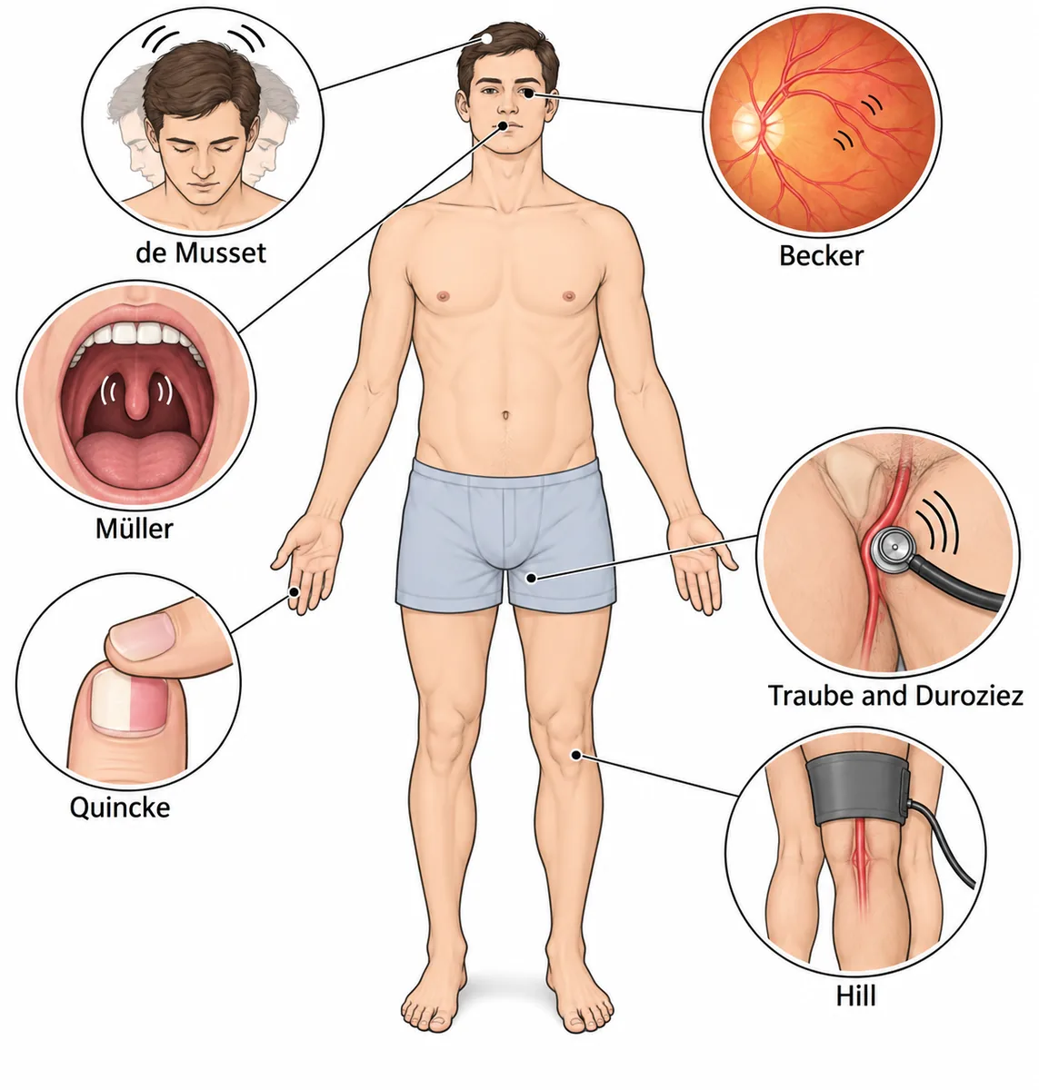
A tall man of 40 with long fingers has a collapsing pulse, a blood pressure of 170/40 mmHg and an early diastolic murmur at the left sternal edge. When his throat is examined, the uvula pulsates with each heartbeat. What is this sign called?
Look first: work out the pulse pressure first, then match each eponym to the place where the throb is seen or heard.
- ATraube sign
- BQuincke sign
- CMüller sign
- Dde Musset sign
Show the answer and explanation
Answer: C. Müller sign
 View larger ↗⤢ Tap to enlarge
C. Müller sign. A pulse pressure of 130 mmHg with an early diastolic murmur is chronic severe aortic regurgitation. The ventricle ejects its normal output plus the regurgitant volume, which drives the systolic pressure up, and the blood falling back in diastole drops the diastolic pressure low. The wide pulse pressure makes the arteries throb wherever they lie near the surface; each eponym simply marks where that throb is seen or heard. Systolic pulsation of the uvula is Müller sign. His tall build and long fingers point to Marfan syndrome and a dilated aortic root as the cause.
Why the other options lose: Traube sign (A) is a pistol-shot sound heard over the femoral artery with each beat. Quincke sign (B) is visible capillary pulsation in the nail bed when the fingertip is gently pressed. de Musset sign (D) is nodding of the head in time with the heartbeat, not movement of the uvula.
Recallab question 61454 · Medicine › Cardiology · reference: Harrison's Principles of Internal Medicine
What the leak does to the left ventricle
Recallab example 2 · a large cavity, not a thick wall
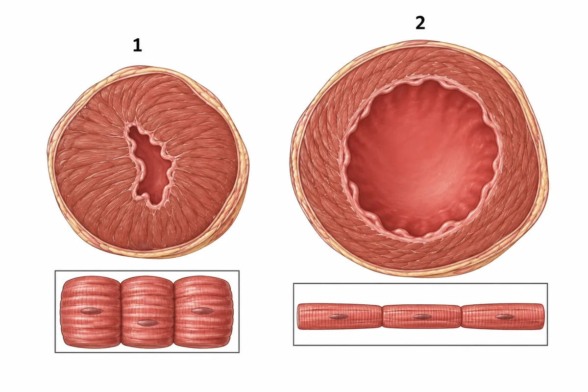
Two left ventricles are drawn in the figure cut across at mid level, each beside a sketch of its muscle fibres. Heart 2 comes from a man with long-standing aortic regurgitation. How were sarcomeres added in it? Two left ventricles cut across at mid level, each beside a sketch of its muscle fibres.
Look first: compare the cavities before the walls, then look at whether the fibres in each inset are wider or longer.
 View larger ↗⤢ Tap to enlarge
- ANot added, existing fibres stretched
- BIn parallel, thickening the fibres
- CIn series, lengthening the fibres
- DReplaced by collagen
Show the answer and explanation
Answer: C. In series, lengthening the fibres
C. In series, lengthening the fibres. Aortic regurgitation returns blood to the ventricle in every diastole, so the ventricle must hold and eject a larger volume. New sarcomeres are laid down end to end, lengthening each fibre; the cavity enlarges and the wall thickens only enough to keep pace. That is eccentric hypertrophy, the pattern of a volume load, and it is heart 2. Heart 1, with a thick wall and a small cavity, is the concentric hypertrophy of a pressure load such as aortic stenosis or long-standing hypertension.
Why the other options lose: Not added, existing fibres stretched (A): the inset shows extra striped segments added end to end, so new sarcomeres have been laid down, not merely stretched. In parallel, thickening the fibres (B) thickens the fibres and the wall; that is heart 1. Replaced by collagen (D): the fibres drawn are lengthened muscle, not scar; collagen replaces muscle after infarction.
Recallab question 56017 · Pathology › CVS · reference: Robbins, Cotran & Kumar Pathologic Basis of Disease
Recallab example 3 · angina with clear coronary arteries
A 55-year-old man in Mumbai has severe chronic aortic regurgitation, exertional angina, BP 158/42 mmHg and left ventricular enlargement. Coronary angiography shows no obstructive disease. Haemoglobin is normal, and he reports mild fatigue. Which principal haemodynamic factor reduces coronary supply?
Look first: ask when the left ventricle fills its own coronary arteries, and what the leak does to the pressure at that moment.
- AReduced aortic valve orifice area
- BReduced left ventricular muscle mass
- CReduced aortic diastolic pressure
- DIncreased coronary vascular resistance
Show the answer and explanation
Answer: C. Reduced aortic diastolic pressure
 View larger ↗⤢ Tap to enlarge
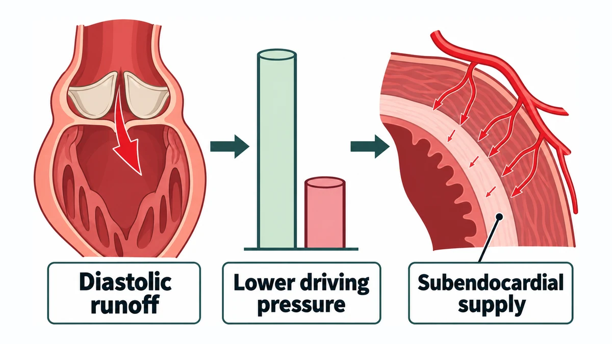
C. Reduced aortic diastolic pressure. The left ventricle is perfused mainly in diastole, and the pressure that drives that flow is the aortic diastolic pressure. Here it is 42 mmHg, because the leak runs blood back into the ventricle through diastole. At the same time the enlarged, hypertrophied ventricle needs more oxygen, and a raised end-diastolic pressure narrows the gradient to the subendocardium further. Exertion raises demand and shortens diastole, so angina appears with clear coronary arteries. With a normal haemoglobin, anaemia does not explain it.
Why the other options lose: Reduced aortic valve orifice area (A) is aortic stenosis, not regurgitation. Reduced left ventricular muscle mass (B) is the wrong way round: chronic severe regurgitation increases the mass and the workload. Increased coronary vascular resistance (D) can influence flow, but the direct limit on supply here is the low upstream diastolic pressure.
Recallab question 74225 · Medicine › Cardiology · reference: Harrison's Principles of Internal Medicine
When the leak is sudden
Recallab example 4 · a new murmur with no wide pulse pressure
A 44-year-old man in a Lucknow hospital has prolonged fever and positive blood cultures. He abruptly develops pulmonary oedema and hypotension with a new short early-diastolic murmur at the left sternal border. Bedside echocardiography shows a normal-sized left ventricle; pulse pressure is not widened. What is the most likely diagnosis?
Look first: weigh the size of the ventricle against the speed of the illness: a dilated ventricle takes time to build.
- AAcute severe aortic regurgitation
- BChronic compensated aortic regurgitation
- CObstructive hypertrophic cardiomyopathy
- DSevere calcific aortic stenosis
Show the answer and explanation
Answer: A. Acute severe aortic regurgitation
 View larger ↗⤢ Tap to enlarge
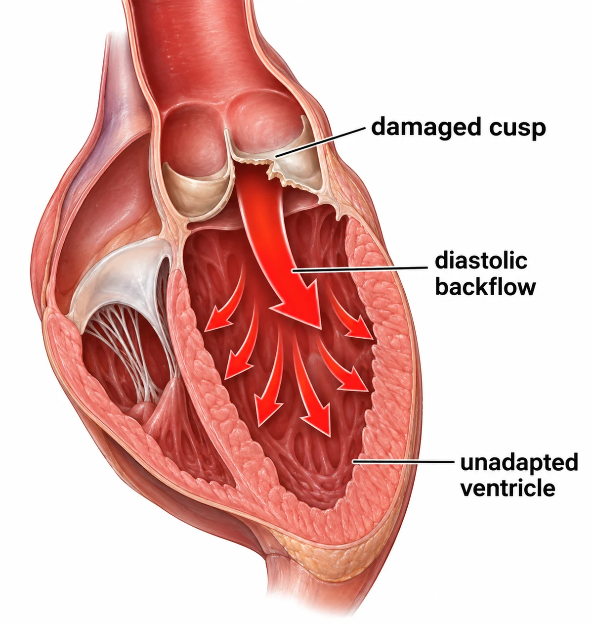
A. Acute severe aortic regurgitation. Infective endocarditis can destroy an aortic cusp and let blood return into the ventricle in diastole all at once. A normal-sized ventricle has not adapted to take that extra volume, so its diastolic pressure rises steeply and the congestion passes back to the lungs. Aortic and ventricular pressures equalise quickly, which shortens the murmur. With low forward output and no chronic adaptation, the expected wide pulse pressure does not develop. The ventricular pressure can even rise above the left atrial pressure and close the mitral valve early. Acute severe regurgitation with haemodynamic compromise needs urgent specialist assessment for valve intervention.
Why the other options lose: Chronic compensated aortic regurgitation (B) gives the ventricle time to enlarge and does not cause this abrupt collapse. Obstructive hypertrophic cardiomyopathy (C) gives a systolic murmur from dynamic outflow obstruction, not a new early diastolic leak. Severe calcific aortic stenosis (D) obstructs outflow in systole, with an ejection murmur rather than an early diastolic one.
Recallab question 72907 · Medicine › Cardiology · reference: Harrison's Principles of Internal Medicine
Why the valve leaks: the root, not the cusps
Recallab example 5 · a stiff spine and a long PR interval
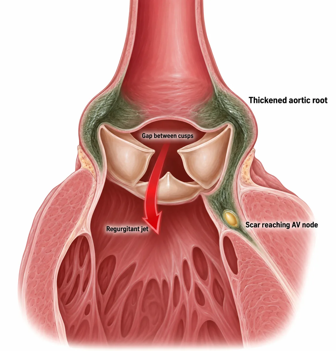
A 50-year-old man has had ankylosing spondylitis for 25 years, with a fused spine and two attacks of anterior uveitis. He is now breathless on exertion. His BP is 150/50 mmHg and his ECG shows a PR interval of 240 ms. Echocardiography is most likely to show which valve lesion?
Look first: put the wide pulse pressure and the long PR interval in one place, just above and just below the aortic valve.
- AMitral stenosis
- BAortic stenosis
- CAortic regurgitation
- DPulmonary regurgitation
Show the answer and explanation
Answer: C. Aortic regurgitation
 View larger ↗⤢ Tap to enlarge
C. Aortic regurgitation. HLA-B27 aortitis inflames the aortic root just above the valve. The root dilates and the cusps thicken and retract, so the valve leaks in diastole and the pulse pressure widens. Its murmur is early diastolic along the sternal edge, heard best with the patient leaning forward; when the root itself is dilated it is often loudest at the right sternal edge. Fibrosis spreading below the valve reaches the AV node and bundle, so a lengthening PR interval, or complete heart block, may accompany the murmur.
Why the other options lose: Mitral stenosis (A) is a rheumatic lesion, not a feature of spondyloarthritis, and it narrows rather than widens the pulse pressure. Aortic stenosis (B) narrows the pulse pressure with a slow-rising pulse; aortitis dilates the root and makes the valve leak, not narrow. Pulmonary regurgitation (D) follows pulmonary hypertension; it does not widen the systemic pulse pressure or involve the conducting system.
Recallab question 61049 · Medicine › Cardiology · reference: Harrison's Principles of Internal Medicine
Recallab example 6 · an aorta like the bark of a tree
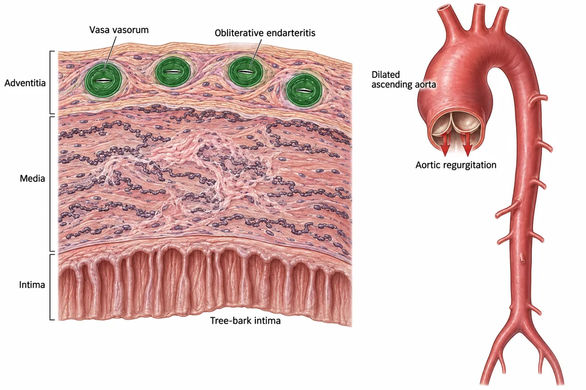
An elderly man has an aneurysm of the ascending aorta and aortic regurgitation. The intimal surface of the aorta shows longitudinal wrinkling like the bark of a tree, and there is obliterative endarteritis of the vasa vasorum. What is the cause?
Look first: ask which layer of the wall is attacked first: the lining, or the small vessels that feed the media.
- AAtherosclerosis
- BTakayasu arteritis
- CTertiary syphilis
- DMarfan syndrome
Show the answer and explanation
Answer: C. Tertiary syphilis
 View larger ↗⤢ Tap to enlarge
C. Tertiary syphilis. Syphilis attacks the vasa vasorum, the small vessels that feed the aortic wall, not the intima. The obliterative endarteritis starves the media, and its elastic tissue scars. The weakened ascending aorta dilates into an aneurysm, dilatation of the valve ring gives aortic regurgitation, and the overlying intima puckers into the tree-bark appearance. The same process can narrow the coronary ostia, so angina can occur with otherwise normal coronary arteries.
Why the other options lose: Atherosclerosis (A) affects the abdominal aorta far more than the ascending aorta, and produces raised lipid plaques rather than tree-bark wrinkling. Takayasu arteritis (B) is a granulomatous large-vessel arteritis of young women that narrows the arch branches rather than causing an ascending aneurysm. Marfan syndrome (D) causes cystic medial degeneration in a young patient, with a dilated sinus and a high risk of dissection.
Recallab question 51219 · Pathology › CVS · reference: Robbins, Cotran & Kumar Pathologic Basis of Disease
The one-minute routine
| If the question gives | Ask first | It settles |
|---|---|---|
| A pulsation named after a person | Where is the throb seen or heard? | Uvula Müller, head de Musset, nails Quincke, groin Traube |
| A large ventricle after a long-standing leak | Were sarcomeres added end to end or side by side? | Volume load adds them in series: a dilated, eccentric ventricle |
| Angina with clear coronary arteries | What is the diastolic pressure? | A low diastolic pressure lowers the coronary driving pressure |
| A new diastolic murmur in endocarditis with shock | Is the ventricle dilated and the pulse pressure wide? | A normal ventricle and pulse pressure mean an acute leak |
| Ankylosing spondylitis with a long PR interval | Is the root inflamed above and below the valve? | Aortitis widens the root and scars the AV node |
| Tree-bark intima in an ascending aneurysm | Which vessels does the disease attack? | Syphilis obliterates the vasa vasorum |
Practise cardiology questions and read the heart's electrical side in ECG questions. For pressure and volume traces, see physiology graphs.
Why candidates use Recallab
- Built towards 22,000 — a batch at a time
- Illustrations drawn for the question
- Every option explained, not just the right one
- AI study tools that are live today
- Start free, today
Or see the plans, or try a timed mock section.
Register to practise NEET PG cardiology questionsFree account, no card needed. Practise with every option explained and a reference on every question.Drawn from questions #61454, #56017, #74225, #72907, #61049, #51219 in the Recallab bank, each with its own verified answer and current-edition reference. Last reviewed 26 October 2026.