
Recallab › Blog › ECG questions in NEET PG: six strips you can read in thirty seconds
ECG questions in NEET PG: six strips you can read in thirty seconds
ECG questions are common in NEET PG, and they are an easy score with focused learning: a small set of patterns, read in the same order every time.
Register to practise NEET PG ECG questions →Free account, no card needed.
An ECG in a question paper looks like a lot of information, and candidates either rush it or skip it. But the question almost always turns on one feature: a pause, an interval, a voltage, the shape of one segment. Learn the patterns that keep coming back and a fixed routine for finding them, and these become marks you can count on. Below are six real Recallab MentorQs, each built on a strip. Try each before you open the answer.
A routine for every strip
- Read what is printed first. A rate, a time bar, a measured amplitude, a bracketed interval. When a figure prints a measurement, use it rather than counting squares.
- Rate and regularity. Fast or slow, regular or irregularly irregular, and whether there is a pause.
- P waves. Is there one before every QRS, and do they all look the same?
- Intervals. PR, QRS width and QT.
- Voltage and axis. Tall or deep complexes, and the direction of the QRS in leads I and aVF.
- ST segment and T wave. Raised, depressed, domed, towering or inverted.
Rate and rhythm
Recallab example 1 · narrow-complex tachycardia
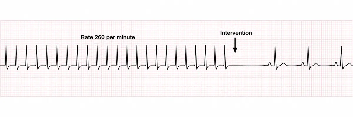
A six-year-old has had a racing heart for an hour; he is alert and pink, with a normal blood pressure. His rhythm strip is drawn in the figure. What should be done first?
Look first: read the printed rate and look for P waves before the complexes, then go back to the stem: how well he is perfused decides the treatment.
 View larger ↗⤢ Tap to enlarge
- AIntravenous verapamil
- BIntravenous adenosine
- CSynchronised DC cardioversion
- DA vagal manoeuvre
Show the answer and explanation
Answer: D. A vagal manoeuvre
Rule: a stable child in supraventricular tachycardia gets a vagal manoeuvre first, then adenosine. Regular narrow complexes at 260 a minute with no visible P waves are re-entry through the AV node; sinus tachycardia does not run this fast in a child. He is alert and well perfused, so there is time for the least invasive step, such as iced water to the face.
Why the closest rival loses: adenosine (B) is the next step if the vagal manoeuvre fails. Cardioversion (C) is for the child who is shocked, and verapamil (A) can cause collapse in young children.
Recallab question 55981 · Paediatrics › Paediatric Cardiology · reference: Ghai Essential Pediatrics
Recallab example 2 · ambulatory recordings
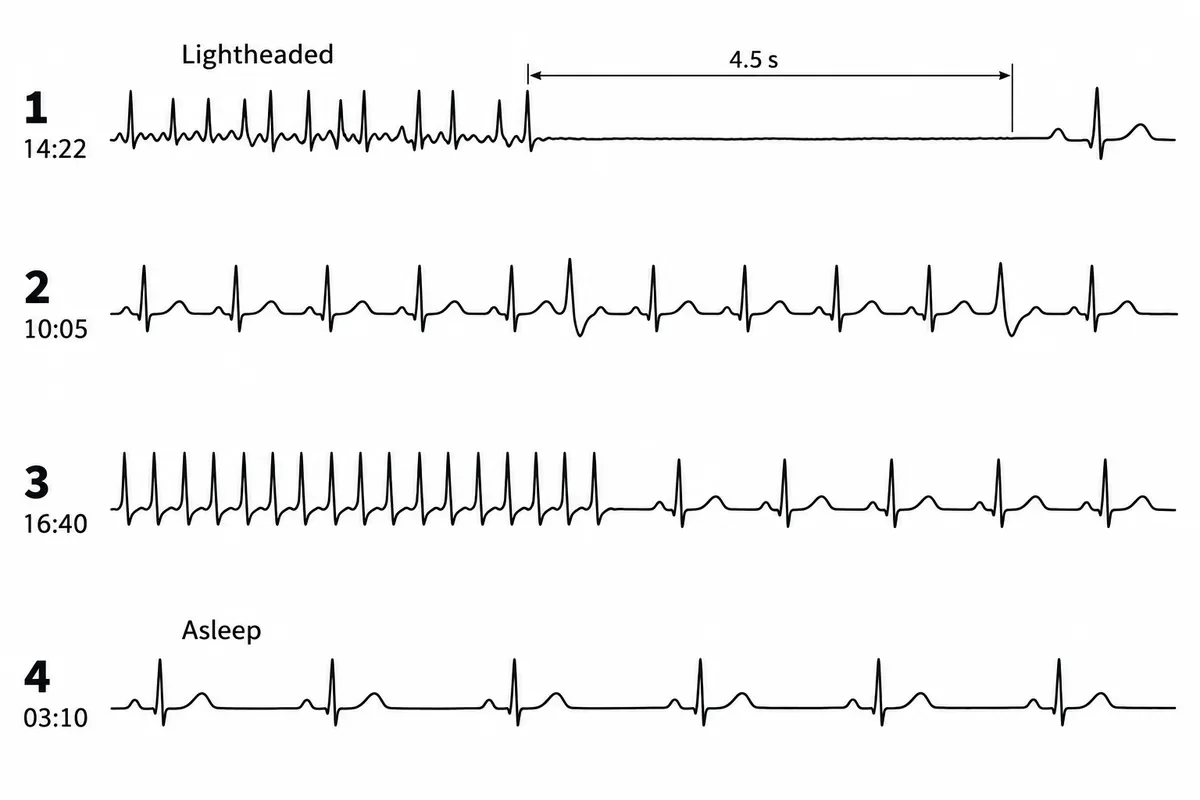
A 72-year-old has dizzy spells and palpitations. Ambulatory recordings from four older patients are drawn in the figure, each with the clock time it was taken. Which recording needs a permanent pacemaker?
Look first: find the strip where a symptom is written, then look at the moment the fast rhythm stops.
 View larger ↗⤢ Tap to enlarge
- ARecording 3
- BRecording 1
- CRecording 2
- DRecording 4
Show the answer and explanation
Answer: B. Recording 1
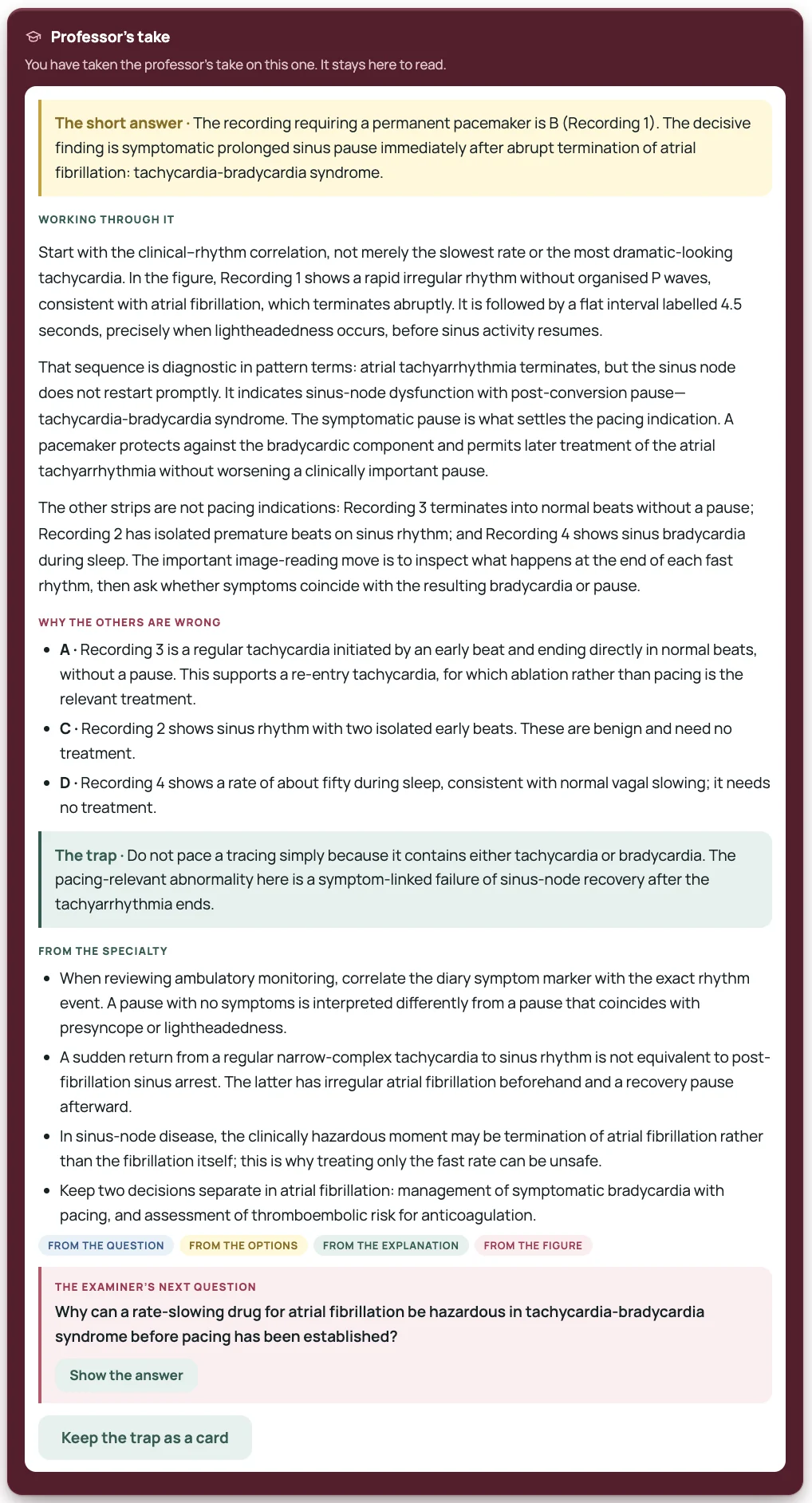
Rule: in tachycardia-bradycardia syndrome the dangerous pause comes as the fast rhythm stops. Recording 1 is atrial fibrillation, irregularly irregular with no P waves, that ends in a 4.5-second pause while he is lightheaded: a diseased sinus node that fails to restart. Drugs to slow the fast runs would lengthen the pauses, so he is paced first and the rate controlled after.
Why the closest rival loses: recording 3 (A) also has a fast run that stops, but normal beats follow at once, with no pause; a regular tachycardia like that is re-entry. Recording 2 (C) is single early beats, and recording 4 (D) is normal slowing during sleep.
Recallab question 57947 · Medicine › Cardiology · reference: Harrison's Principles of Internal Medicine
Intervals: the QT
Recallab example 3 · audiogram and rhythm strip
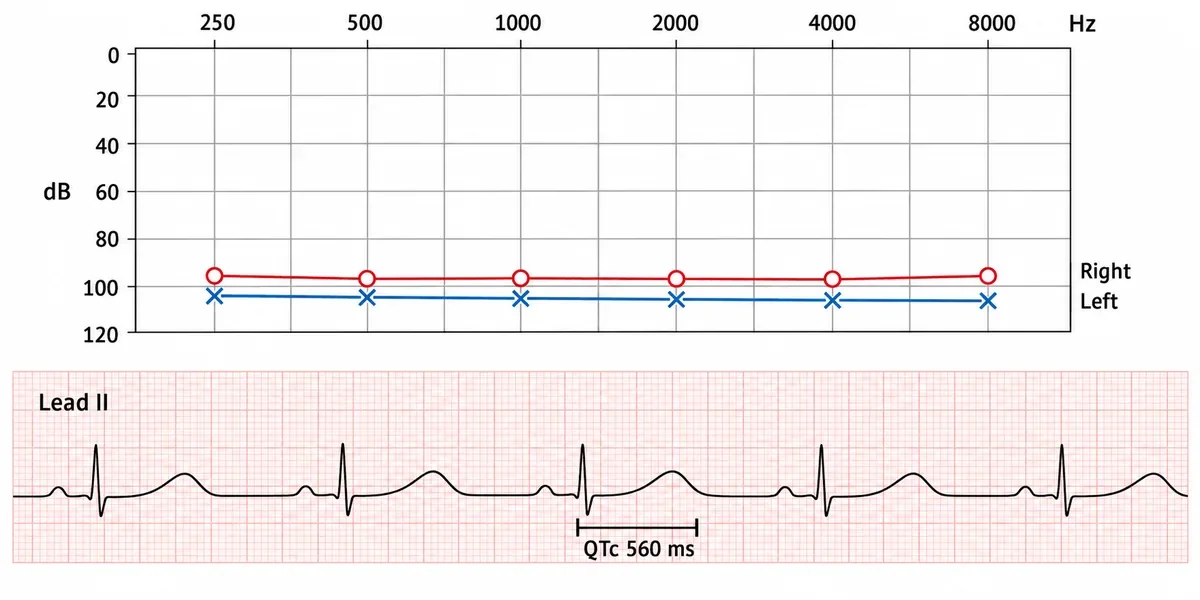
A boy of six, deaf from birth, has twice collapsed briefly: once when a dog jumped up at him and once while swimming. His parents are first cousins and both hear normally. His hearing test and a strip of his resting ECG are drawn in the figure. What is the diagnosis?
Look first: read both panels before the options: the audiogram gives the deafness, the bracket gives the QT.
 View larger ↗⤢ Tap to enlarge
- APendred syndrome
- BJervell and Lange-Nielsen syndrome
- CRomano-Ward syndrome
- DUsher syndrome
Show the answer and explanation
Answer: B. Jervell and Lange-Nielsen syndrome
Rule: deafness from birth with a long QT interval is Jervell and Lange-Nielsen syndrome. The audiogram shows profound loss in both ears at every frequency, and the corrected QT of 560 ms is far above normal. Related parents who hear normally fit recessive inheritance, and a fright or swimming is the adrenergic trigger of a dangerous ventricular rhythm; a beta blocker is started.
Why the closest rival loses: Romano-Ward syndrome (C) is also a long QT, but dominant and with normal hearing. Pendred (A) pairs deafness with a goitre and Usher (D) with retinitis pigmentosa; neither lengthens the QT.
You can check the figure: this strip is drawn to scale. The QT spans about 0.66 s and the beats are about 1.35 s apart, and 0.66 ÷ √1.35 is about 0.57 s, which is the printed QTc.
Recallab question 57588 · ENT › Diseases of the ear · reference: Dhingra, Diseases of Ear, Nose and Throat
Recallab example 4 · three lead II strips
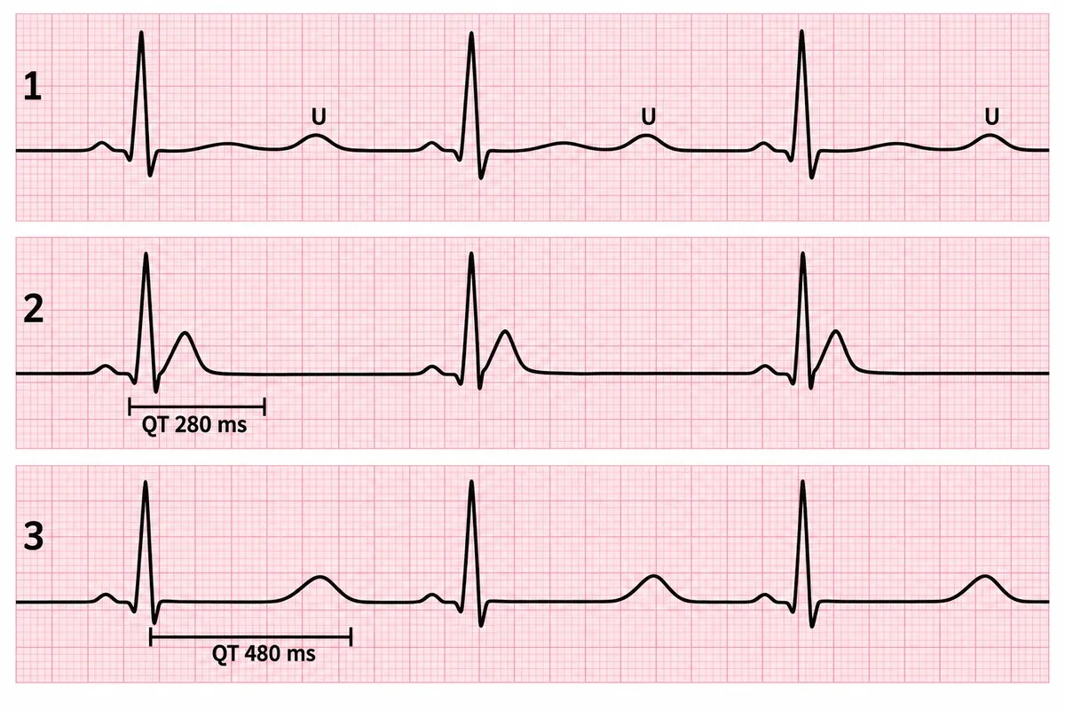
A woman with lung cancer is confused, constipated and dry. Three lead II strips from patients with different electrolyte disturbances are drawn in the figure, and one of them matches her. What is given first?
Look first: name her electrolyte from the stem first, then use the printed QT values and see which segment makes the difference.
 View larger ↗⤢ Tap to enlarge
- AIntravenous furosemide
- BIntravenous calcium gluconate
- CIntravenous saline
- DIntravenous potassium
Show the answer and explanation
Answer: C. Intravenous saline
Rule: a high calcium shortens the QT by shortening the ST segment. Confusion, constipation and dehydration in lung cancer are hypercalcaemia, and strip 2, with almost no ST segment, is hers. Hypercalcaemia causes polyuria, so she is volume depleted; intravenous saline comes first.
Why the closest rival loses: calcium gluconate (B) treats a low calcium, the long flat ST segment of strip 3. Potassium (D) is for strip 1, whose flat T waves and U waves are a low potassium. Furosemide (A) before rehydration deepens the volume loss.
Recallab question 56752 · Medicine › Electrolytes · reference: Harrison's Principles of Internal Medicine
Voltage and the ST segment
Recallab example 5 · voltage
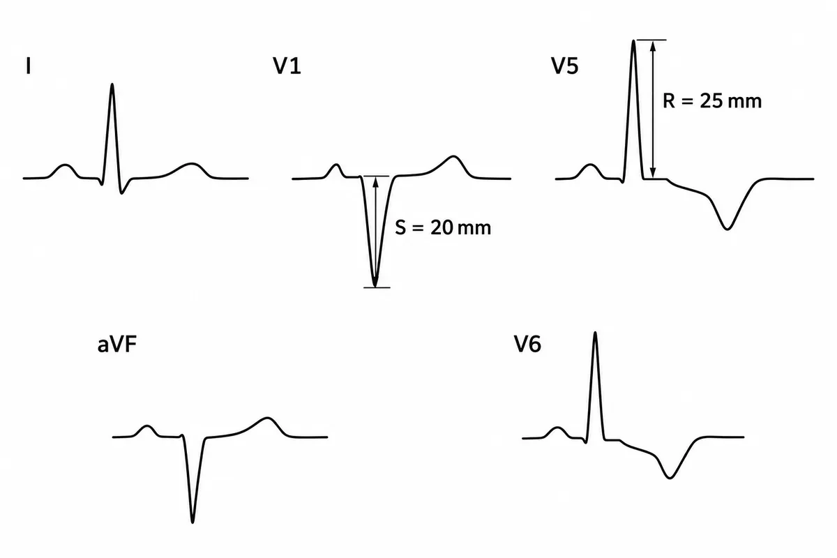
The electrocardiogram of a man with long-standing hypertension is drawn in the figure, with two amplitudes measured. What does the tracing show?
Look first: add the two measured amplitudes before you look at the ST segment.
 View larger ↗⤢ Tap to enlarge
- ARight ventricular hypertrophy with strain
- BLeft ventricular hypertrophy with strain
- CLeft bundle branch block
- DLateral ischaemia alone
Show the answer and explanation
Answer: B. Left ventricular hypertrophy with strain
Rule: S in V1 plus R in V5 or V6 above 35 mm is left ventricular hypertrophy (Sokolow-Lyon). Here 20 + 25 = 45 mm. Downsloping ST depression with asymmetric T inversion in V5 and V6 is the strain pattern of a thick, pressure-loaded wall.
Why the closest rival loses: left bundle branch block (C) needs a broad QRS, and these are narrow. Right ventricular hypertrophy (A) gives a dominant R in V1 and a right axis; here V1 is a deep S and the axis is to the left. Ischaemia alone (D) does not explain the voltage.
Recallab question 55271 · Medicine › Cardiology · reference: Harrison's Principles of Internal Medicine
Recallab example 6 · infarct in time
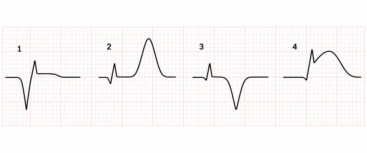
A man of 58 arrived within half an hour of crushing central chest pain. One chest lead was recorded repeatedly over the next three days, and four of those complexes are drawn in the figure out of order. Which complex was recorded first?
Look first: put all four in order before you answer: the first has no Q wave and no ST shift, only a changed T wave.
 View larger ↗⤢ Tap to enlarge
- AComplex 3
- BComplex 2
- CComplex 1
- DComplex 4
Show the answer and explanation
Answer: B. Complex 2
Rule: the hyperacute T wave comes first. Within minutes of occlusion the T wave grows tall and broad (complex 2). The ST segment then lifts into a dome (4), Q waves form over hours (1), and over days the ST segment settles and the T wave inverts (3). The order is 2, 4, 1, 3.
Why the closest rival loses: complex 4 (D) is the classic picture of an infarct, which is why it tempts, but its ST elevation follows the hyperacute T. A deep Q wave (C) takes hours, and T inversion (A) belongs to the following days.
Recallab question 56147 · Medicine › Cardiology · reference: Harrison's Principles of Internal Medicine
For the complex strips: ask for a Professor’s take
Some strips carry more than one idea. Example 2 asks you to compare four recordings against a symptom; example 6 asks you to put four complexes in order. For questions like these, answer first, then request a Professor’s take: a structured review of the answer you chose, with the verdict, the reasoning, the alternatives, the trap and a follow-up question. Use it to check how you read the strip, then close it and read the next strip on your own.
 View larger ↗⤢ Tap to enlarge
The thirty-second routine
| Step | Look at | It usually settles |
|---|---|---|
| 1. What is printed | Rate, time bar, measured amplitudes, brackets | The numbers the question wants you to use |
| 2. Rate and regularity | Fast or slow, regular or not, any pause | Tachycardia, fibrillation, sick sinus |
| 3. P waves | One before every QRS, all the same shape? | Sinus rhythm, re-entry, fibrillation |
| 4. Intervals | PR, QRS width, QT | Block, bundle branch block, long or short QT |
| 5. Voltage and axis | S in V1 plus R in V5 or V6; leads I and aVF | Hypertrophy and axis deviation |
| 6. ST and T | Raised, depressed, domed, towering, inverted | Infarction and its age, strain |
Other posts in this series: six biostatistics chart questions and ten NEET PG image questions, from heart-sound traces to slit-lamp views. For reading what a question asks, see NEET PG question types.
For the potassium and sodium results behind many ECG changes, see six NEET PG acid-base and electrolyte questions.
Why candidates use Recallab
- Built towards 22,000 — a batch at a time
- Illustrations drawn for the question
- Every option explained, not just the right one
- AI study tools that are live today
- Start free, today
Or see the plans, or try a timed mock section.
Register to practise NEET PG ECG questionsFree account, no card needed. Practise with every option explained and a reference on every question.Drawn from questions #55981, #57947, #57588, #56752, #55271, #56147 in the Recallab bank, each with its own verified answer and current-edition reference. Last reviewed 25 September 2026.