
Recallab › Blog › Acid-base and electrolytes for NEET PG: six lab pictures and the number to read first
Acid-base and electrolytes for NEET PG: six lab pictures and the number to read first
Electrolyte questions look like arithmetic, but each one hides a single question: where did the ion go, or where did the result come from?
Register to practise NEET PG medicine questions →Free account, no card needed.
These questions show a lab card, a chart or a column of ions and ask what explains it. The pictures below are illustrations drawn for the questions, not scans of reports. Each is a real Recallab MentorQ, and each answer turns on one feature of the picture. Try each before you open the answer.
Before you read the numbers: three questions
- Is the result real? A sample, a tube or a site can distort one value without changing the patient.
- Which route explains it? Gut, kidney, intake or a drug: the urine and the history usually decide.
- Which number comes first? The anion gap, the measured osmolality or the urine potassium often locates the cause before the cause is named.
Acid and base
Recallab example 1 · the anion gap
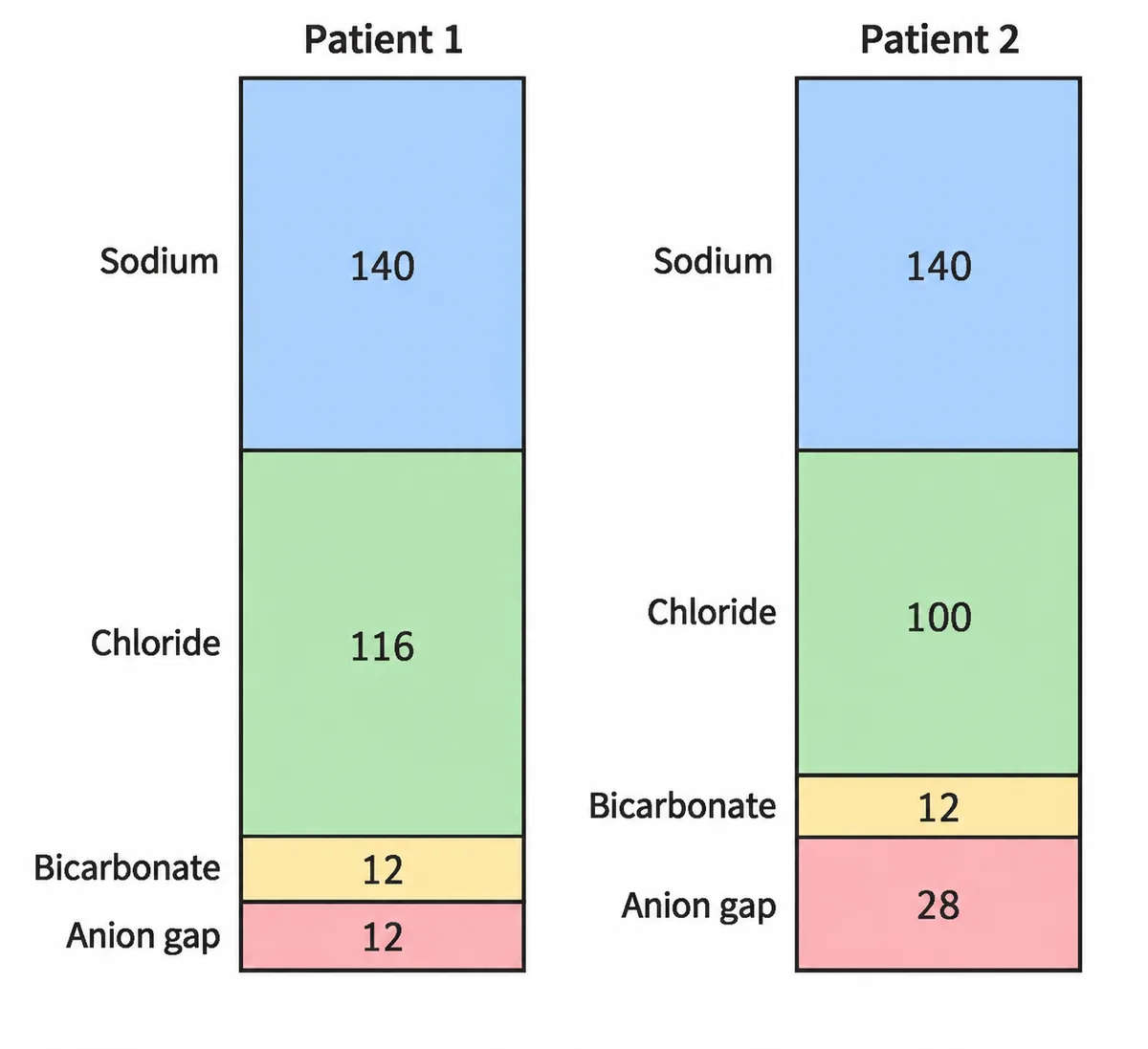
Two columns of blood ion values are drawn in the figure for two patients with low bicarbonate, each labelled with sodium, chloride, bicarbonate and the unmeasured anion gap. Patient 1's chloride is expanded; patient 2's anion gap is expanded instead. Which patient has diarrhoea?
Look first: ask which block has grown, and what that block says about where the bicarbonate went.
 View larger ↗⤢ Tap to enlarge
- APatient 2, high anion gap acidosis
- BPatient 1, hyperchloraemic acidosis
- CPatient 2, hyperchloraemic acidosis
- DPatient 1, high anion gap acidosis
Show the answer and explanation
Answer: B. Patient 1, hyperchloraemic acidosis
Patient 1, hyperchloraemic acidosis. Diarrhoeal stool carries bicarbonate out, which lowers plasma bicarbonate directly. The body retains chloride to balance the charge, so chloride rises while the anion gap stays normal: exactly patient 1's column. Renal tubular acidosis gives the same normal anion gap picture, and the urinary anion gap helps tell the two apart.
Why the closest rival loses: patient 1 with a high anion gap (D) is the right patient with the wrong reading: patient 1's chloride is raised, not the gap. Patient 2 (A and C) has a widened gap and a normal chloride, which points to an unmeasured acid such as ketones. Chloride up, gap normal: diarrhoea. Gap up, chloride normal: an unmeasured acid.
Recallab question 55348 · Medicine › Electrolyte · reference: Harrison's Principles of Internal Medicine
Recallab example 2 · the blood gas sample
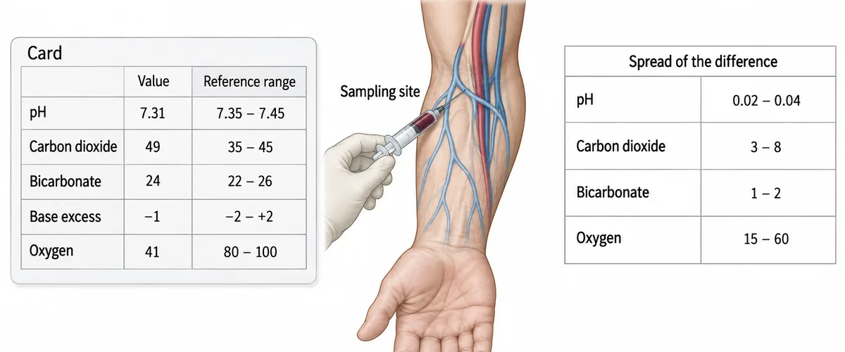
A breathless adult has had a full gas panel measured. The issued card is drawn in the figure beside a note of where the sample came from and a small table of how far each measured quantity is known to differ between the two sampling sites. Which of the printed numbers may be acted on?
Look first: find the sampling site first, then look for the row in the table whose spread is wide.
 View larger ↗⤢ Tap to enlarge
- AThe whole panel
- BNone of the panel
- COxygen row after correction
- DThe acid-base rows only
Show the answer and explanation
Answer: D. The acid-base rows only
The acid-base rows only. The needle is in a superficial vein, not an artery. A peripheral venous sample tracks arterial acidity, carbon dioxide and bicarbonate closely enough to judge acid-base: the table shows their differences are narrow. Its oxygen is what one tissue bed left behind and has no fixed relation to the arterial value: the oxygen difference spans a wide range, so no single figure can be recovered from it.
Why the closest rival loses: acting on the whole panel (A) ignores the evidence in the table that one row behaves differently from the rest. Acting on none of it (B) throws away a usable acid-base reading and costs the patient a second needle. Correcting the oxygen row (C) does not work, because the table gives a range, not a constant. Venous blood answers the acid question; only an artery answers the oxygen one.
Recallab question 59012 · Biochemistry › Acid-Base & Clinical Biochemistry · reference: Harper's Illustrated Biochemistry
Sodium
Recallab example 3 · a low sodium that is not real
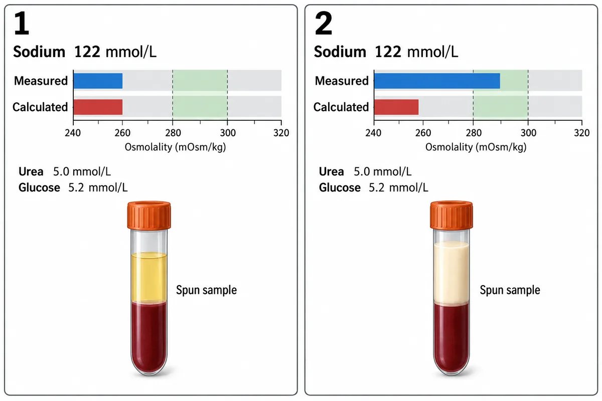
Two patients each have a serum sodium of 122 millimoles per litre. The figure sets out for each of them the osmolality measured by the laboratory, the osmolality calculated from the sodium, urea and glucose, and the appearance of the sample after spinning. What explains the low sodium in patient 2?
Look first: compare the measured and the calculated osmolality on each card, then look at the spun tube.
 View larger ↗⤢ Tap to enlarge
- APsychogenic polydipsia
- BPseudohyponatraemia from hyperlipidaemia
- CInappropriate antidiuretic hormone secretion
- DHyperglycaemic hyponatraemia
Show the answer and explanation
Answer: B. Pseudohyponatraemia from hyperlipidaemia
Pseudohyponatraemia from hyperlipidaemia. The calculated osmolality starts from the sodium, so it follows a low sodium down. The measured value is independent of it and depends on real particles. In patient 2 the measured value is normal, so the cells see a normal tonicity, and the creamy plasma layer shows what is displacing the water in that sample. In patient 1 the two values agree, so that sodium is genuinely low and must be treated.
Why the closest rival loses: inappropriate antidiuretic hormone secretion (C) and psychogenic polydipsia (A) are true dilutional states, which lower the measured osmolality; patient 2's is normal. Hyperglycaemic hyponatraemia (D) is out because the glucose is normal and a glucose load would raise the calculated value as well. Measure it before you treat it.
Recallab question 57255 · Medicine › Electrolyte · reference: Harrison's Principles of Internal Medicine
Recallab example 4 · a low sodium from what they eat
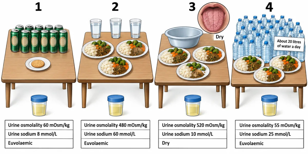
Four adults each have a serum sodium of about 118 mmol/L. What each takes in on a typical day is drawn in the figure, with their urine results and fluid status. Which patient's low sodium comes from too little dietary solute?
Look first: read each table for what goes in, then each card for how dilute the urine is.
 View larger ↗⤢ Tap to enlarge
- APatient 4
- BPatient 3
- CPatient 1
- DPatient 2
Show the answer and explanation
Answer: C. Patient 1
Patient 1. The kidney needs solute to excrete water. Urine can be diluted to about 50 to 100 mOsm/kg and no further, so the water cleared in a day is capped by the solute taken in that day. Beer supplies water and carbohydrate but very little salt or protein. Eating almost nothing, he can clear only a few litres before water is retained. His urine is maximally dilute and almost free of sodium. When food or saline restores solute, free water pours out and the sodium can climb too fast.
Why the closest rival loses: patient 4 (A) has equally dilute urine but eats normal meals; twenty litres a day of water overwhelms even a full solute intake. Patient 3 (B) is dry with concentrated urine and a low urine sodium, which is volume depletion. Patient 2 (D) has concentrated, sodium-rich urine despite a low sodium and a normal diet, which is inappropriate antidiuresis. No solute in, no water out.
Recallab question 57852 · Medicine › Electrolyte · reference: Harrison's Principles of Internal Medicine
Potassium
Recallab example 5 · a low potassium and its route
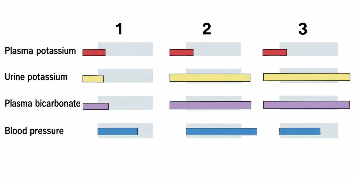
Three patients all have a plasma potassium of 2.6 millimoles per litre. Their urine potassium, plasma bicarbonate and blood pressure are drawn in the figure as bars against their normal bands. Patient 1 is losing potassium through the gut. What in the figure proves it?
Look first: start with the plasma potassium (the same in all three), then ask what a working kidney does when it is low.
 View larger ↗⤢ Tap to enlarge
- AA low bicarbonate
- BA low urine potassium
- CA high bicarbonate
- DA high blood pressure
Show the answer and explanation
Answer: B. A low urine potassium
A low urine potassium. A kidney that is working conserves potassium hard once the plasma level falls. Patient 1's urine potassium is low while the plasma level is low, which is that conservation, so the loss must be elsewhere, and the gut is the only other large route. Patients 2 and 3 are still pouring potassium into the urine, so the kidney is the leak.
Why the closest rival loses: a low bicarbonate (A) is present in patient 1 and fits intestinal fluid losses, but it only narrows the cause; the urine potassium locates the route. A high bicarbonate (C) belongs to patients 2 and 3, whose losses are renal. A high blood pressure (D) goes with patient 2's mineralocorticoid excess, a renal loss. Low in, low out means the kidney is innocent.
Recallab question 56451 · Medicine › Electrolyte · reference: Harrison's Principles of Internal Medicine
Recallab example 6 · a high potassium and the drug that started it
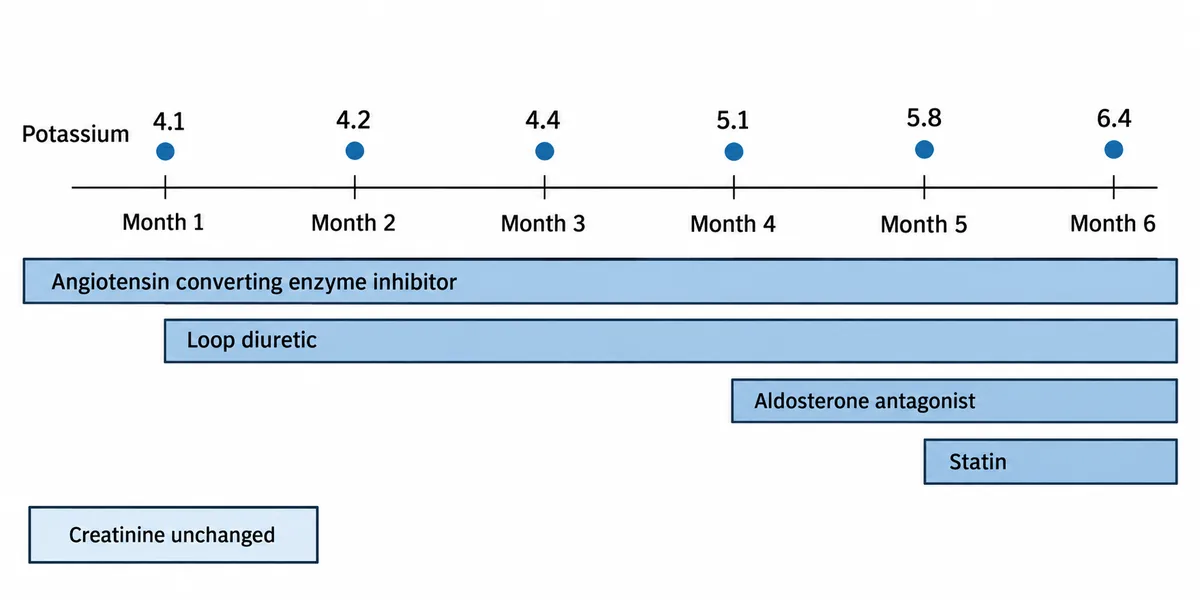
A woman with heart failure and stable kidney function is admitted with a potassium of 6.4 millimoles per litre. The figure sets her potassium over the past six months beside the dates on which each of her drugs was started. Which change explains the rise?
Look first: find the month where the potassium turns, then see what started in that month.
 View larger ↗⤢ Tap to enlarge
- AStarting the loop diuretic
- BStarting the enzyme inhibitor
- CStarting the aldosterone antagonist
- DA fall in kidney function
Show the answer and explanation
Answer: C. Starting the aldosterone antagonist
Starting the aldosterone antagonist. Aldosterone drives sodium reabsorption and potassium secretion in the collecting duct, and blocking its receptor removes the main route by which potassium leaves the body. The first three values are flat and the climb begins at the fourth month, which is when this drug was started; no other bar starts there. When a result and a prescription change on the same date, the prescription is the first suspect.
Why the closest rival loses: the enzyme inhibitor (B) does raise potassium, but it was running unchanged through three normal results. The loop diuretic (A) was started two months earlier and lowers potassium. A fall in kidney function (D) is excluded by the box recording an unchanged creatinine. Line the results up against the prescription dates.
Recallab question 56352 · Medicine › Electrolyte · reference: Harrison's Principles of Internal Medicine
The one-minute routine
| If you see | Read first | It usually settles |
|---|---|---|
| Columns of ions | Which block has grown: chloride or the anion gap | Where the bicarbonate went |
| A gas card and a sampling site | Which row is wide in the table | Which numbers can be acted on |
| A low sodium with two osmolalities | Do the measured and calculated values agree | Real or artefact |
| A low sodium and what they eat | How dilute is the urine, and what solute goes in | Intake, water or hormone |
| A low potassium with urine bars | Is the urine potassium low or high | Gut or kidney |
| A high potassium and a timeline | Which drug starts where the line turns | The drug, not the kidney |
For other kinds of figure, see ten NEET PG image questions and six eye and ENT spotters. For the decision that follows the picture, see next-best-step management questions.
Why candidates use Recallab
- Built towards 22,000 — a batch at a time
- Illustrations drawn for the question
- Every option explained, not just the right one
- AI study tools that are live today
- Start free, today
Or see the plans, or try a timed mock section.
Register to practise NEET PG medicine questionsFree account, no card needed. Practise with every option explained and a reference on every question.Drawn from questions #55348, #59012, #57255, #57852, #56451, #56352 in the Recallab bank, each with its own verified answer and current-edition reference. Last reviewed 9 October 2026.