
Recallab › Blog › Medicine for NEET PG: six rheumatology figures, from joints to systemic clues
Medicine for NEET PG: six rheumatology figures, from joints to systemic clues
A rheumatology figure becomes easier when you read its distribution first. Which joints, which part of a cell or which vessel is involved? The pattern connects the picture to the patient.
Register to practise NEET PG Medicine questions →Free account, no card needed.
These six illustrated Recallab questions move from joint disease to its wider clinical setting. Start with hand and sacroiliac patterns, then follow the clues into antibody testing, limb pressures and gastric bleeding. Whether you are learning Medicine for the first time or revising for NEET PG or INI-CET, try each question before opening its worked answer.
Before you choose: three questions
- Where is the change? Name the joints or anatomical region involved, and check which neighbouring structures are spared.
- What has changed? Separate soft-tissue swelling, erosion, sclerosis and new bone; in a fluorescence field or vessel drawing, describe the visible pattern before naming a disease.
- Which clinical clue fits the picture? Match the distribution to the timing of pain, antibody result, skin finding or pressure difference. Then check whether the question asks for a diagnosis, a stage or a treatment.
Three joint patterns: fingers, hand margins and sacroiliac joints
Recallab example 1 · a whole finger and its nail
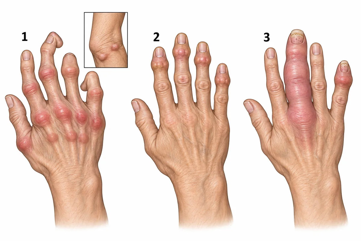
Three hands are drawn in the figure with the swollen joints shaded and the nails shown. Which two features in the drawing mark the hand with psoriatic arthritis? Three hands with the swollen joints shaded and the nails drawn.
Look first: compare the swollen joints, then check whether the swelling extends between them and whether the nails change.
 View larger ↗⤢ Tap to enlarge
- ADistal joint involvement alone
- BSymmetrical knuckles and elbow nodules
- CSausage finger and nail pitting
- DBony lumps at the fingertip joints
Show the answer and explanation
Answer: C. Sausage finger and nail pitting
C. Sausage finger and nail pitting. Hand 3 has dactylitis: the whole digit is swollen, including the tissue between its joints. Its nails are pitted and lift away at the free edge, a change called onycholysis. Together these findings identify psoriatic arthritis. The asymmetrical distribution and involvement of a joint near the fingertip support the same diagnosis. Look for the combination rather than relying on the joint site alone.
Why the other options lose: distal joint involvement alone (A) also occurs in osteoarthritis, so it does not distinguish the two. Symmetrical knuckles and elbow nodules (B), drawn in hand 1, fit rheumatoid arthritis. Bony lumps at the fingertip joints (D), drawn in hand 2 without nail changes or whole-digit swelling, fit osteoarthritis.
Recallab question 55162 · Medicine › Immunology & Rheumatology · reference: Harrison's Principles of Internal Medicine
Recallab example 2 · the margins of the hand joints
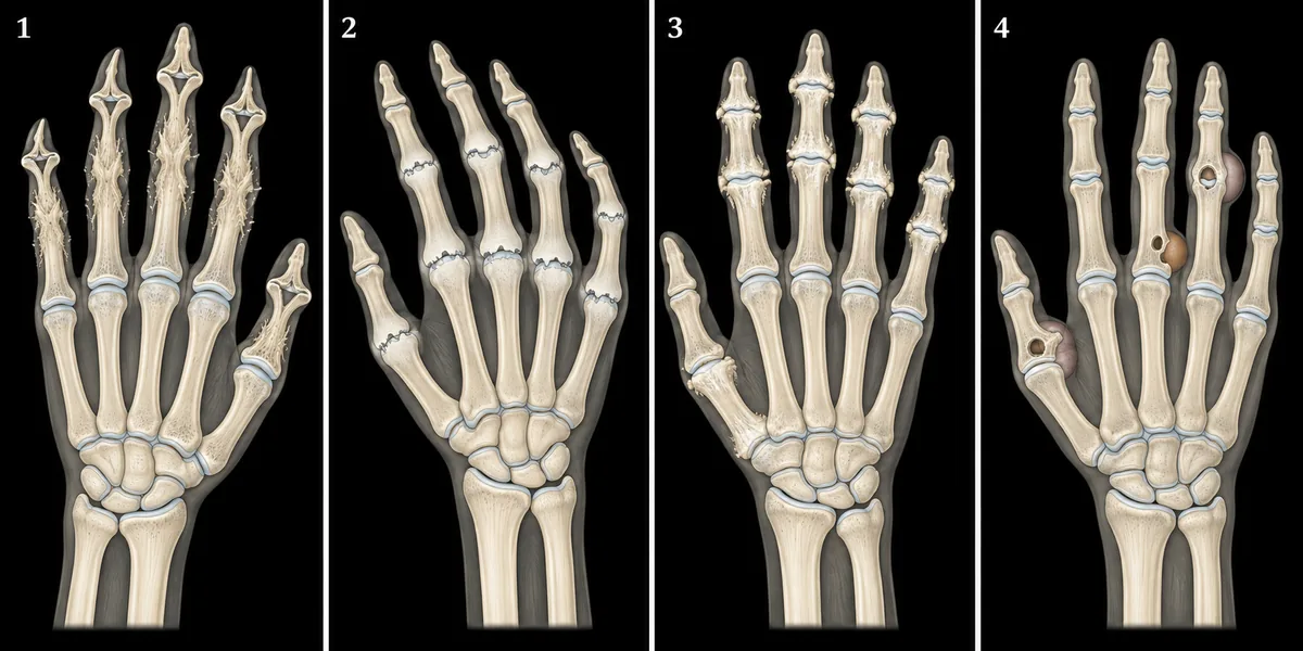
A 42-year-old woman has had painful, stiff hands every morning for a year, and her anti-cyclic citrullinated peptide (anti-CCP) antibody is strongly positive. Four hand radiographs are drawn in the figure. Which hand is hers? Four hand radiographs drawn at one scale, numbered 1 to 4.
Look first: start with the knuckles and middle finger joints; compare the erosion edges, nearby bone density and fingertip joints.
 View larger ↗⤢ Tap to enlarge
- AHand 1
- BHand 4
- CHand 2
- DHand 3
Show the answer and explanation
Answer: C. Hand 2
C. Hand 2. The knuckle and middle finger joints have uniform narrowing, marginal erosions and thinning of the nearby bone, called periarticular osteopenia. The fingertip joints are spared. This is the rheumatoid pattern that fits prolonged morning stiffness and a strongly positive anti-CCP antibody. Inflamed synovium erodes the unprotected bone at the joint margins. Anti-CCP supports the diagnosis and is associated with a greater likelihood of erosive disease.
Why the other options lose: hand 1 (A) has pencil-in-cup changes at the fingertip joints with new bone formation, fitting psoriatic arthritis. Hand 4 (B) has punched-out erosions with overhanging edges and soft-tissue lumps, fitting gout. Hand 3 (D) has osteophytes and subchondral sclerosis at the fingertip and middle finger joints and the thumb base, fitting osteoarthritis. Their erosion shapes and distributions do not match the rheumatoid pattern.
Recallab question 55947 · Medicine › Immunology & Rheumatology · reference: Harrison's Principles of Internal Medicine
Recallab example 3 · the first definite sacroiliac change
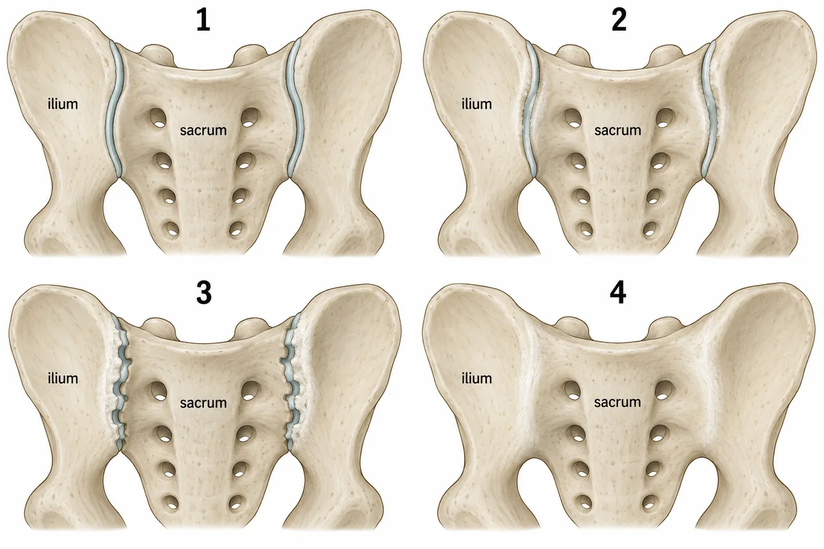
A man of twenty-eight has had eight months of low back pain that wakes him in the second half of the night and eases once he moves about. Four possible appearances of a pair of sacroiliac joints are drawn in the figure as cropped pelvic radiographs. Which pair shows the earliest change that counts as definite sacroiliitis? Four pelvic radiographs cropped to both sacroiliac joints, drawn at one scale.
Look first: compare the joint margins before judging the width: distinguish a blurred edge, a notch in the bone and a complete bony bridge.
 View larger ↗⤢ Tap to enlarge
- APair 2
- BPair 3
- CPair 1
- DPair 4
Show the answer and explanation
Answer: B. Pair 3
B. Pair 3. Both joints show erosions with adjacent sclerosis. The erosions cut notches into the subchondral bone, making parts of the space appear wider; the dense band beside them is sclerosis. These are definite structural changes, unlike blurring alone. The clinical pattern also fits inflammatory back pain: waking late at night and improving with movement. The question asks for the earliest definite appearance among these drawings, so do not jump to the most advanced image.
Why the other options lose: pair 2 (A) has indistinct margins alone, a suspicious finding rather than definite radiographic sacroiliitis. Pair 1 (C) is normal, with crisp margins and even joint spaces. Pair 4 (D) shows complete fusion, which is definite disease but a late stage. MRI can show inflammation before definite changes appear on a plain radiograph.
Recallab question 57455 · Medicine › Immunology & Rheumatology · reference: Harrison's Principles of Internal Medicine
Beyond the joints: antibodies, arteries and the stomach
Recallab example 4 · skin thickening and a fluorescence field
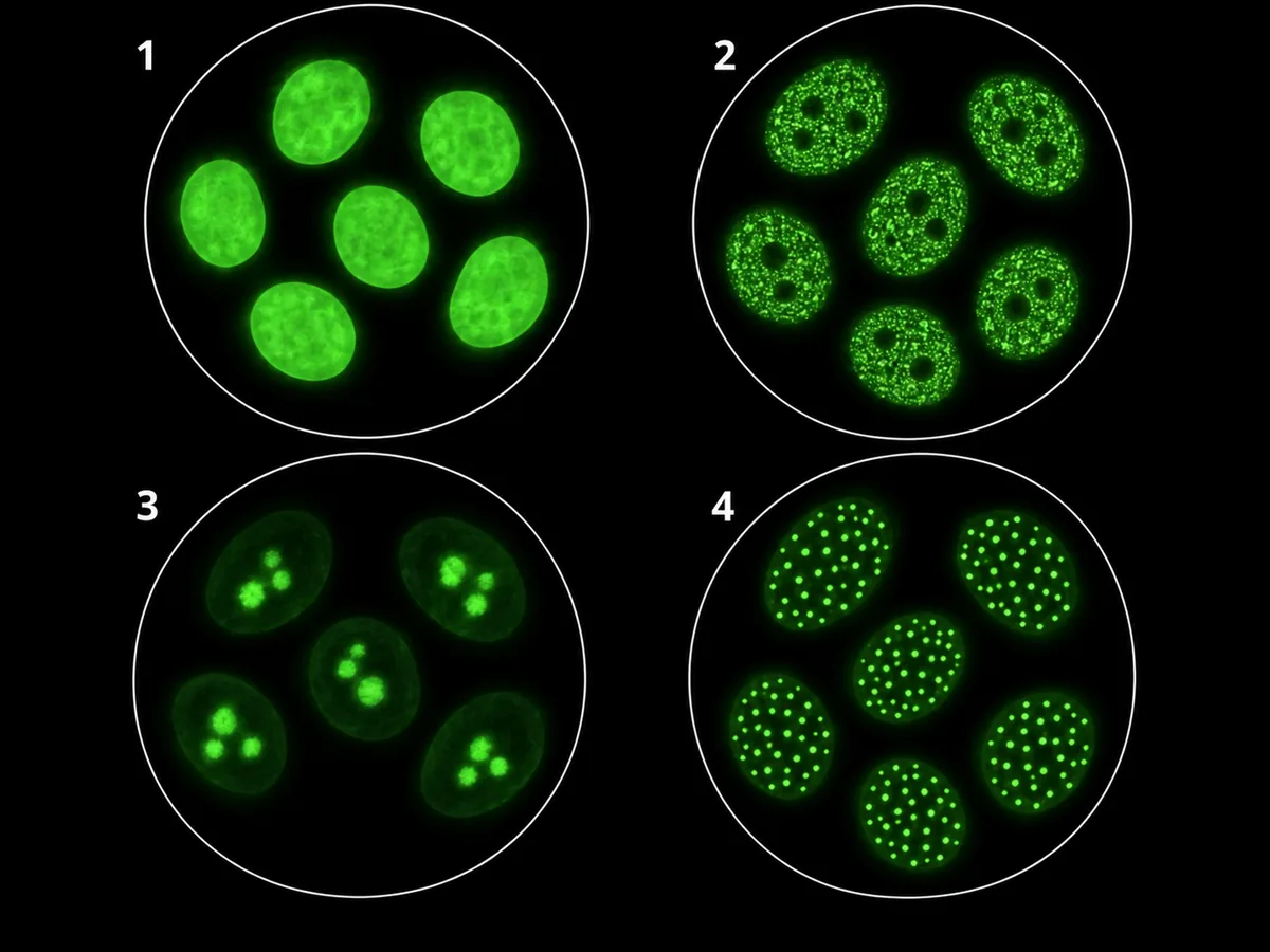
A woman with Raynaud phenomenon has skin thickening that has spread in a year from her fingers onto her forearms and trunk. Four antinuclear antibody patterns on HEp-2 cells are drawn in the figure. Which pattern fits her? Four antinuclear antibody patterns on HEp-2 cells, numbered 1 to 4.
Look first: decide how far the skin thickening extends, then compare whether fluorescence fills the nucleus, forms speckles or is confined to smaller structures.
 View larger ↗⤢ Tap to enlarge
- AField 1, homogeneous
- BField 3, nucleolar
- CField 4, centromere
- DField 2, speckled
Show the answer and explanation
Answer: B. Field 3, nucleolar
B. Field 3, nucleolar. The spread of skin thickening onto the trunk points to diffuse cutaneous systemic sclerosis. Field 3 lights up the nucleoli while the rest of each nucleus remains dim. A nucleolar antinuclear antibody pattern is associated with systemic sclerosis and is the best fit among these choices. It is a pointer, so the specific antibody still needs confirmation; the fluorescence pattern alone does not establish the diagnosis or subtype.
Why the other options lose: field 1, homogeneous (A), stains the nuclei evenly and is associated with antibodies to double-stranded DNA or histones, as in lupus. Field 4, centromere (C), shows discrete dots and is more typical of limited cutaneous systemic sclerosis. Field 2, speckled (D), is associated with antibodies to extractable nuclear antigens, as in lupus and mixed connective tissue disease; it is less fitting than the nucleolar pattern in this stem.
Recallab question 55564 · Medicine › Immunology & Rheumatology · reference: Harrison's Principles of Internal Medicine
Recallab example 5 · an arm pressure that misleads
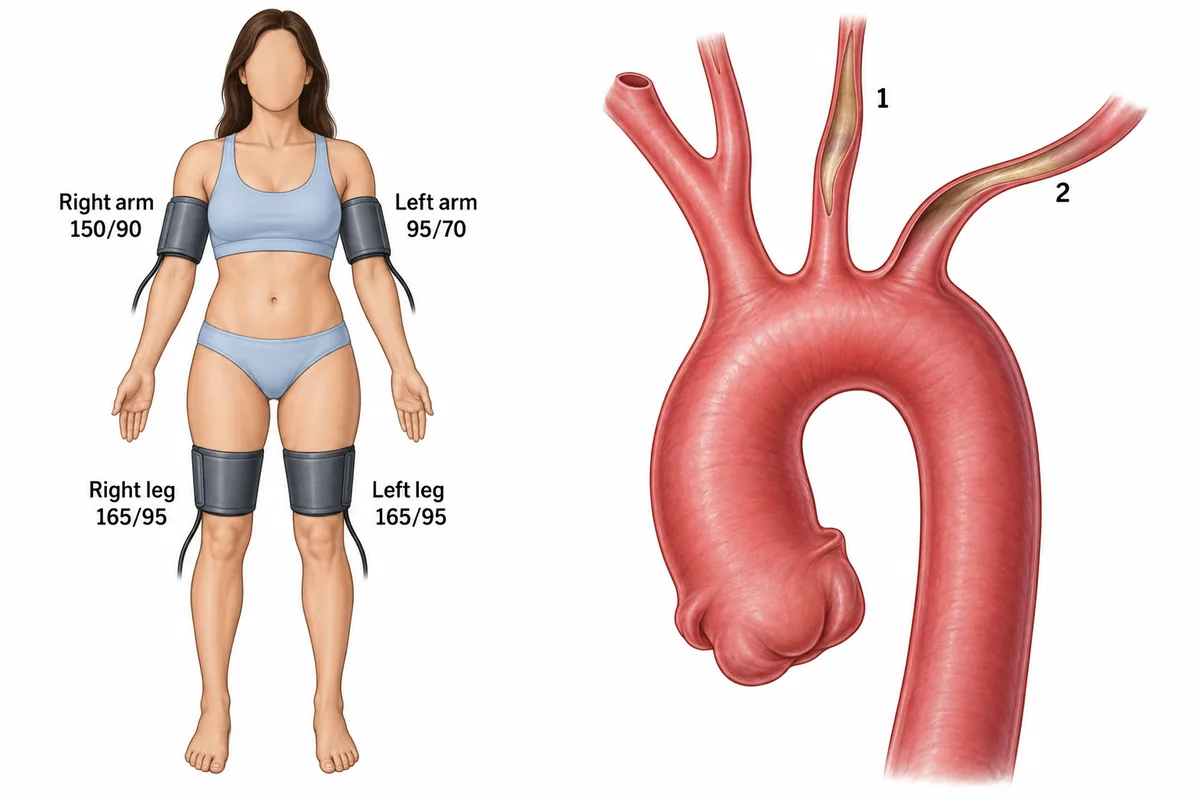
A woman of twenty-six has aching in her left arm when she uses it. Her blood pressure in all four limbs and a drawing of her aortic arch are shown in the figure. What is the diagnosis? Blood pressure in four limbs beside a drawing of the aortic arch and its branches.
Look first: compare all four pressures, then trace the branch supplying the arm with the lower reading.
 View larger ↗⤢ Tap to enlarge
- ATakayasu arteritis
- BThoracic outlet syndrome
- CAortic dissection
- DCoarctation of the aorta
Show the answer and explanation
Answer: A. Takayasu arteritis
A. Takayasu arteritis. A young woman with pain on using her arm and long, smooth narrowing at the aortic arch branch origins fits large-vessel vasculitis. The narrowed left subclavian artery explains both the arm symptoms and the low left-arm reading. The right arm and legs have raised pressures: the left-arm value underestimates the pressure because blood reaches that cuff through a narrowed artery. Follow blood pressure in a limb with an open supplying artery rather than accepting the lowest value.
Why the other options lose: thoracic outlet syndrome (B) causes positional compression and does not explain narrowing at the arch branch origins. Aortic dissection (C) usually presents suddenly with pain and an intimal flap, which is absent from the drawing. Coarctation of the aorta (D) typically produces higher arm than leg pressures with delayed femoral pulses; here the leg pressures are higher, and the drawing shows branch narrowing rather than a focal aortic narrowing.
Recallab question 56760 · Medicine › Immunology & Rheumatology · reference: Harrison's Principles of Internal Medicine
Recallab example 6 · gastric bleeding with tight skin
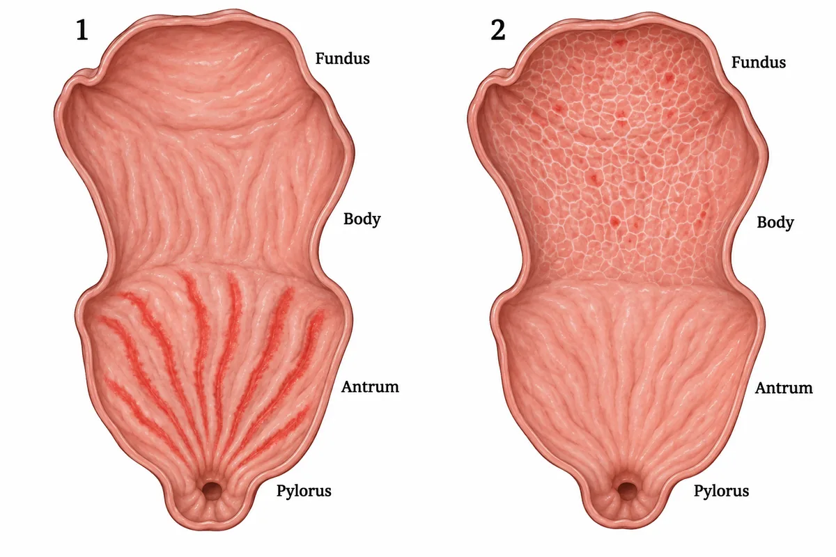
Two patients with slow bleeding from the stomach and iron deficiency had upper endoscopy, drawn in the figure. One is a woman with tight, thickened skin over her fingers and face; the other is a man with large varices at the lower oesophagus. How is the woman's lesion treated? Two stomachs drawn as seen at endoscopy, the antrum and pylorus at the bottom of each drawing.
Look first: locate the abnormal vessels: compare the antrum near the pylorus with the fundus and upper body.
 View larger ↗⤢ Tap to enlarge
- AIron replacement alone
- BArgon plasma coagulation
- CA transjugular portosystemic shunt
- DA non-selective beta blocker
Show the answer and explanation
Answer: B. Argon plasma coagulation
B. Argon plasma coagulation. The woman has the antral stripes in stomach 1: gastric antral vascular ectasia, often called watermelon stomach. Her tight skin supports its association with systemic sclerosis. Dilated mucosal vessels bleed slowly and cause iron deficiency. Argon plasma coagulation treats those vessels at endoscopy and may need more than one session. The site and pattern distinguish this lesion from the portal hypertensive gastropathy illustrated in stomach 2.
Why the other options lose: iron replacement alone (A) replaces the iron lost but leaves the bleeding vessels untreated. A transjugular portosystemic shunt (C) lowers portal pressure and can treat portal hypertensive gastropathy; it does not treat this antral vascular ectasia. A non-selective beta blocker (D) also lowers portal pressure, which is not the mechanism driving the woman's lesion. Her lesion needs treatment directed at the mucosal vessels.
Recallab question 57157 · Medicine › Gastroenterology · reference: Harrison's Principles of Internal Medicine
The one-minute routine
| If the figure shows | Ask first | It points towards |
|---|---|---|
| A whole swollen digit with altered nails | Are there pits or lifting at the nail edge? | Psoriatic arthritis when these features occur together |
| Marginal hand erosions with nearby bone thinning | Are the knuckles and middle finger joints involved, with the tips spared? | Rheumatoid arthritis |
| Irregular sacroiliac margins | Is there erosion with sclerosis, or only blurring? | Definite radiographic sacroiliitis when erosions and sclerosis are present |
| Fluorescence confined to nucleoli | Does the skin thickening extend onto the trunk? | Systemic sclerosis; confirm the specific antibody |
| One low arm pressure with narrowed arch branches | What do the other arm and legs read? | Takayasu arteritis; assess pressure in a limb with an open artery |
| Antral stripes in a patient with tight skin | Are the vessels near the pylorus or in the fundus? | Gastric antral vascular ectasia; endoscopic treatment |
For more figure practice, see ten NEET PG image questions and physiology graphs. For the association between systemic sclerosis and gastric antral vascular ectasia, see the SCOT trial endoscopy study.
Why candidates use Recallab
- Built towards 22,000 — a batch at a time
- Illustrations drawn for the question
- Every option explained, not just the right one
- AI study tools that are live today
- Start free, today
Or see the plans, or try a timed mock section.
Register to practise NEET PG Medicine questionsFree account, no card needed. Practise with every option explained and a reference on every question.Drawn from questions #55162, #55947, #57455, #55564, #56760, #57157 in the Recallab bank, each with its own verified answer and current-edition reference. Last reviewed 14 October 2026.