
Recallab › Blog › Physiology for NEET PG: six figures from ion gradients to muscle contraction
Physiology for NEET PG: six figures from ion gradients to muscle contraction
Follow the ions before naming the response. Pumps establish gradients, channels use them, and calcium links electrical signals to transmitter release and contraction.
Register to practise NEET PG physiology questions →Free account, no card needed.
These six illustrated Recallab questions trace that chain from the cell membrane to the muscle filament. They connect first-year MBBS physiology with NEET PG and INI-CET revision for interns and graduates. Try each question before opening its worked answer, and keep track of which side of which membrane each ion occupies.
Before you follow the ions: three questions
- Which membrane and compartment? Separate the cell surface, nerve terminal and intracellular calcium store.
- What drives the movement? Look for ATP use, an ion gradient or a channel opening towards its reversal potential.
- Which event comes next? Distinguish transmitter release, calcium storage and cross-bridge detachment rather than treating contraction as one step.
Pumps, nerve conduction and inhibitory signals
Recallab example 1 · phosphate on the pump
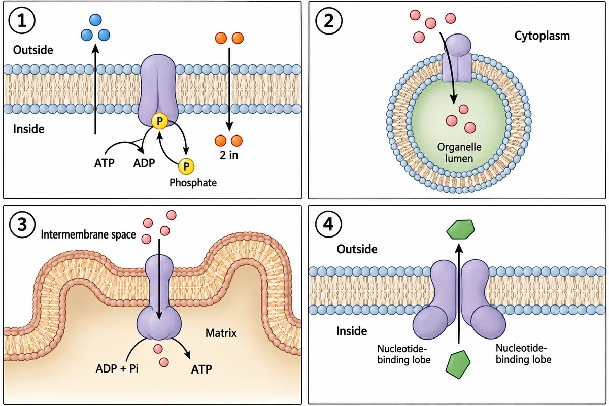
Four pumps are drawn in the figure, each in the membrane where it belongs and each with what it moves. Which numbered pump works by attaching a phosphate group to itself during each cycle, and what family does that put it in? Four pumps drawn in the membranes where they belong, with what each one moves.
Look first: check whether phosphate attaches to the protein itself, then count the ions moving each way.
 View larger ↗⤢ Tap to enlarge
- APump 2, a V-type pump
- BPump 4, an ATP-binding cassette pump
- CPump 1, a P-type pump
- DPump 3, an F-type pump
Show the answer and explanation
Answer: C. Pump 1, a P-type pump
C. Pump 1, a P-type pump. The defining feature is a phosphorylated intermediate: phosphate attaches to the pump protein during its cycle. Panel 1 shows the sodium-potassium pump moving three sodium ions out and two potassium ions in. Attachment and removal of phosphate drive changes in its shape. ATP use alone does not identify the family; look for phosphate on the protein.
Why the other options lose: Pump 2, a V-type pump (A), acidifies an organelle through a rotary mechanism without forming a phosphorylated pump intermediate. Pump 4, an ATP-binding cassette pump (B), uses nucleotide-binding domains to drive transport, without attaching phosphate to itself. Pump 3, an F-type pump (D), uses the mitochondrial proton gradient to make ATP and also lacks that phosphorylated intermediate.
Recallab question 58609 · Physiology › General Physiology · reference: Ganong's Review of Medical Physiology
Recallab example 2 · the last hump to arrive
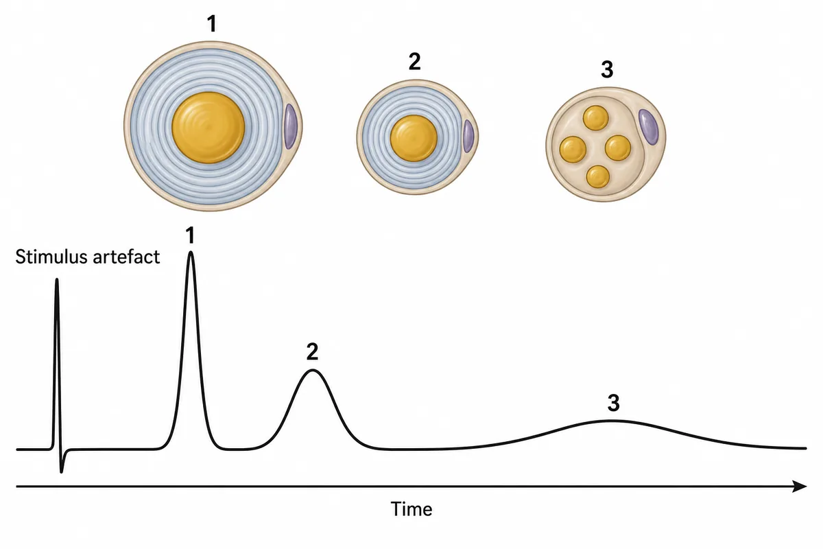
A compound action potential recorded from a mixed nerve is drawn in the figure with three separate humps arriving at different times. Which fibres produced hump 3? One recording from a mixed nerve, three humps and nothing named.
Look first: compare the delay and width of each hump, then relate arrival time to fibre size and myelination.
 View larger ↗⤢ Tap to enlarge
- AUnmyelinated C fibres
- BMyelinated A beta fibres
- CMyelinated A alpha fibres
- DMyelinated A delta fibres
Show the answer and explanation
Answer: A. Unmyelinated C fibres
A. Unmyelinated C fibres. The recording combines signals from populations of fibres travelling at different speeds. Hump 3 arrives last and is broadest, identifying the slow-conducting C fibres. Their small diameter and absence of myelin explain the delay; variation in their arrival times spreads out the hump. Read the time axis before assigning the fibre type.
Why the other options lose: Myelinated A beta fibres (B) conduct faster than C fibres and carry touch signals, so they cannot account for the latest hump. Myelinated A alpha fibres (C) are the largest and fastest fibres here, contributing to the earliest hump. Myelinated A delta fibres (D) are thinner and more lightly myelinated than the fastest fibres, but still conduct faster than C fibres; they fit the intermediate hump.
Recallab question 55009 · Physiology › Nerve & Muscle · reference: Ganong's Review of Medical Physiology
Recallab example 3 · find the flat trace
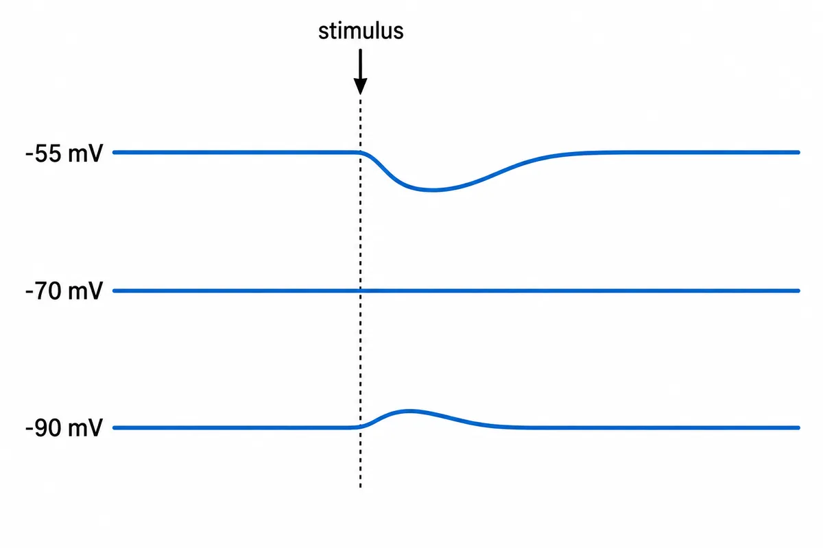
An inhibitory input to a spinal interneuron is stimulated while the cell is held by current injection at three different membrane potentials. In this cell the potassium equilibrium potential is -90 mV and the sodium equilibrium potential is +55 mV. The three recordings are drawn in the figure. Channels for which ion does the transmitter open? Three recordings from one neuron held at three membrane potentials, the same inhibitory input stimulated at the arrow each time.
Look first: find the holding potential at which stimulation produces no voltage deflection, then compare the directions on either side.
 View larger ↗⤢ Tap to enlarge
- ACalcium
- BPotassium
- CChloride
- DSodium
Show the answer and explanation
Answer: C. Chloride
C. Chloride. The response is flat at -70 mV, downward at -55 mV and upward at -90 mV. Its reversal potential is therefore about -70 mV: opening the channels moves the membrane towards that value from either side. This fits chloride, as in fast inhibition through glycine or GABA-A receptors. An upward voltage deflection can still accompany inhibition; increased conductance can shunt excitatory current.
Why the other options lose: Calcium (A) has a strongly positive equilibrium potential and would produce depolarisation at all three holding potentials. Potassium (B) would reverse at the stated -90 mV, making the bottom trace flat rather than upward. Sodium (D) would reverse at the stated +55 mV and also depolarise the cell at all three values. The flat trace identifies the reversal potential more reliably than the direction of one response alone.
Recallab question 69127 · Physiology › Nervous System · reference: Ganong's Review of Medical Physiology
Calcium handling, transmitter release and the cross-bridge
Recallab example 4 · sodium changes calcium extrusion
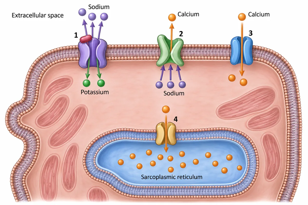
A cardiac myocyte is drawn with four membrane transport proteins numbered. Partial inhibition of numbered protein 1 raises intracellular sodium and eventually strengthens the heartbeat. Which protein carries out the step that links the rise in sodium to the rise in contractile calcium? A cardiac myocyte with four membrane transport proteins numbered and arrows of ion movement.
Look first: follow the sodium gradient from the partly inhibited protein to the protein that moves calcium across the surface membrane.
 View larger ↗⤢ Tap to enlarge
- AProtein 1, the sodium-potassium pump
- BProtein 2, the sodium-calcium exchanger
- CProtein 4, the sarcoplasmic reticulum calcium pump
- DProtein 3, the L-type calcium channel
Show the answer and explanation
Answer: B. Protein 2, the sodium-calcium exchanger
B. Protein 2, the sodium-calcium exchanger. Partial inhibition of protein 1 raises intracellular sodium, reducing the inward sodium gradient. Protein 2 normally uses that gradient to bring sodium in while moving calcium out. With less driving force, it extrudes less calcium. More calcium remains available inside the cell and can be stored in the sarcoplasmic reticulum for the next beat, increasing contraction strength. This is the indirect mechanism of cardiac glycosides such as digoxin.
Why the other options lose: Protein 1, the sodium-potassium pump (A), starts the sequence by changing sodium transport, but it does not transport calcium. Protein 4, the sarcoplasmic reticulum calcium pump (C), stores calcium already within the cell; it does not link the weakened surface sodium gradient to reduced calcium extrusion. Protein 3, the L-type calcium channel (D), permits calcium entry during the plateau, but direct stimulation of that channel is not the mechanism shown.
Recallab question 56908 · Physiology › Cardio Vasular Physiology · reference: Ganong's Review of Medical Physiology
Recallab example 5 · larger responses after the train
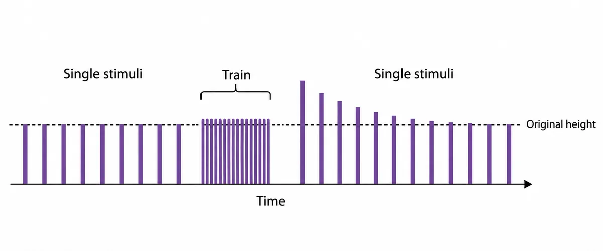
The size of the end-plate potential recorded from one muscle fibre is drawn in the figure as a row of bars, for single stimuli to its motor nerve before and after a brief high-frequency train. What has built up during the train to change the later responses? End-plate potentials from one fibre as a row of bars, before and after a brief high-frequency train.
Look first: compare the first single response after the train with the original height, then follow its gradual return.
 View larger ↗⤢ Tap to enlarge
- APotassium outside the muscle fibre
- BAcetylcholine receptors at the end-plate
- CCalcium in the motor nerve terminal
- DCalcium in the sarcoplasmic reticulum
Show the answer and explanation
Answer: C. Calcium in the motor nerve terminal
C. Calcium in the motor nerve terminal. During the high-frequency train, calcium enters the presynaptic terminal faster than it can be cleared. Residual calcium increases vesicle fusion with subsequent impulses, so each releases more packets of acetylcholine. The end-plate potentials are initially larger, then return towards baseline as calcium is removed. This is post-tetanic potentiation: the relevant calcium is in the nerve terminal.
Why the other options lose: Potassium outside the muscle fibre (A) would depolarise the muscle and reduce the driving force for the end-plate current, tending to shrink the response. Acetylcholine receptors at the end-plate (B) cannot increase in number and disappear again on this brief timescale; structural changes take much longer. Calcium in the sarcoplasmic reticulum (D) affects muscle contraction, whereas these bars measure the electrical end-plate response to transmitter release.
Recallab question 56710 · Physiology › Nerve & Muscle · reference: Ganong's Review of Medical Physiology
Recallab example 6 · letting go before re-cocking
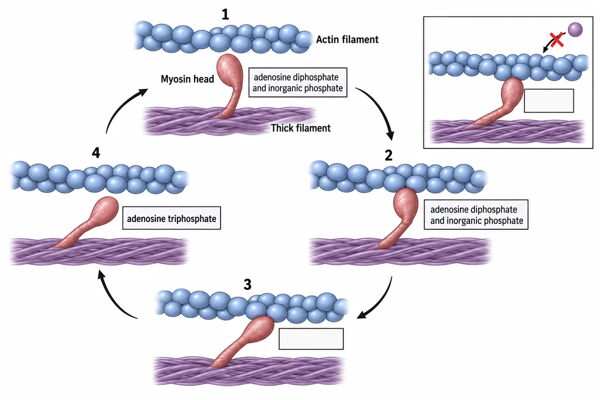
One myosin head and its actin filament are drawn in the figure at four numbered positions around a single cycle, with the nucleotide bound at each position written in a box beside the head. The head lets go of actin between two of the positions. What makes it let go? One myosin head and its actin filament at four numbered positions around a cycle.
Look first: compare the bound nucleotide before and after detachment, then locate the separate re-cocking step.
 View larger ↗⤢ Tap to enlarge
- ABinding of adenosine triphosphate
- BRelease of inorganic phosphate
- CHydrolysis of adenosine triphosphate
- DCalcium binding to troponin
Show the answer and explanation
Answer: A. Binding of adenosine triphosphate
A. Binding of adenosine triphosphate. At position 3, the head is attached to actin with an empty nucleotide-binding site. At position 4, ATP is bound and the head has detached. Binding ATP breaks the actin-myosin attachment; hydrolysis follows and re-cocks the detached head. Without a fresh supply of ATP, heads remain attached, explaining the rigid state in the side panel.
Why the other options lose: Release of inorganic phosphate (B) drives the power stroke while the head remains attached. Hydrolysis of adenosine triphosphate (C) re-cocks a head that has already detached; it is a later event than ATP binding. Calcium binding to troponin (D) exposes binding sites on actin so myosin can attach, rather than releasing an attached head. Keep the sequence clear: ATP binding detaches; ATP hydrolysis re-cocks.
Recallab question 57307 · Physiology › Nerve & Muscle · reference: Ganong's Review of Medical Physiology
The one-minute routine
| If the figure gives | Ask first | It settles |
|---|---|---|
| Four membrane pumps | Does phosphate attach to the protein itself? | A phosphorylated intermediate identifies a P-type pump |
| Three humps in a compound nerve recording | Which arrives last and spreads widest? | Slow unmyelinated C fibres produce the late broad hump |
| Synaptic responses at three holding potentials | Which trace is flat? | Reversal near -70 mV fits chloride here |
| Partial sodium-pump inhibition in a cardiac cell | Which transporter uses the inward sodium gradient to extrude calcium? | Less exchange-driven calcium extrusion increases available calcium |
| Larger end-plate potentials after a train | Which compartment retains calcium briefly? | Residual nerve-terminal calcium increases transmitter release |
| A cross-bridge cycle with nucleotide boxes | What binds when the head detaches? | ATP binding detaches; hydrolysis re-cocks |
Practise physiology questions and build your curve-reading routine with physiology graphs. For other illustrations, see ten NEET PG image questions.
Why candidates use Recallab
- Built towards 22,000 — a batch at a time
- Illustrations drawn for the question
- Every option explained, not just the right one
- AI study tools that are live today
- Start free, today
Or see the plans, or try a timed mock section.
Register to practise NEET PG physiology questionsFree account, no card needed. Practise with every option explained and a reference on every question.Drawn from questions #58609, #55009, #69127, #56908, #56710, #57307 in the Recallab bank, each with its own verified answer and current-edition reference. Last reviewed 23 October 2026.