
Recallab › Blog › The ulnar nerve for NEET PG: why a low lesion claws more than a high one
The ulnar nerve for NEET PG: why a low lesion claws more than a high one
A claw hand is not the nerve's damage; it is the balance of the muscles that are left. Work out which muscles still pull, and the level of the lesion follows.
Register to practise NEET PG anatomy questions →Free account, no card needed.
Clawing is unopposed long extensor action at the knuckles, with flexion of the joints beyond them. The bending of the end joints comes from the ulnar half of flexor digitorum profundus, which acts on the ring and little fingers. That is why the ulnar paradox catches people: the higher, worse lesion looks milder. These six illustrated Recallab questions build the ulnar nerve from the posture of one finger to the timing of a needle study, for NEET PG and INI-CET revision. Try each question before opening its worked answer.
Before you name the level: three questions
- Which muscles still pull? The long extensors keep working when the small hand muscles fail; the posture is the balance of what is left.
- Is profundus spared? A lesion at the wrist leaves the ulnar half of flexor digitorum profundus working; a lesion at the elbow takes it too.
- Which branch, and when? In the palm the nerve splits by job, and a needle study only shows denervation weeks after the injury.
The claw and the paradox
Recallab example 1 · one finger adrift at rest
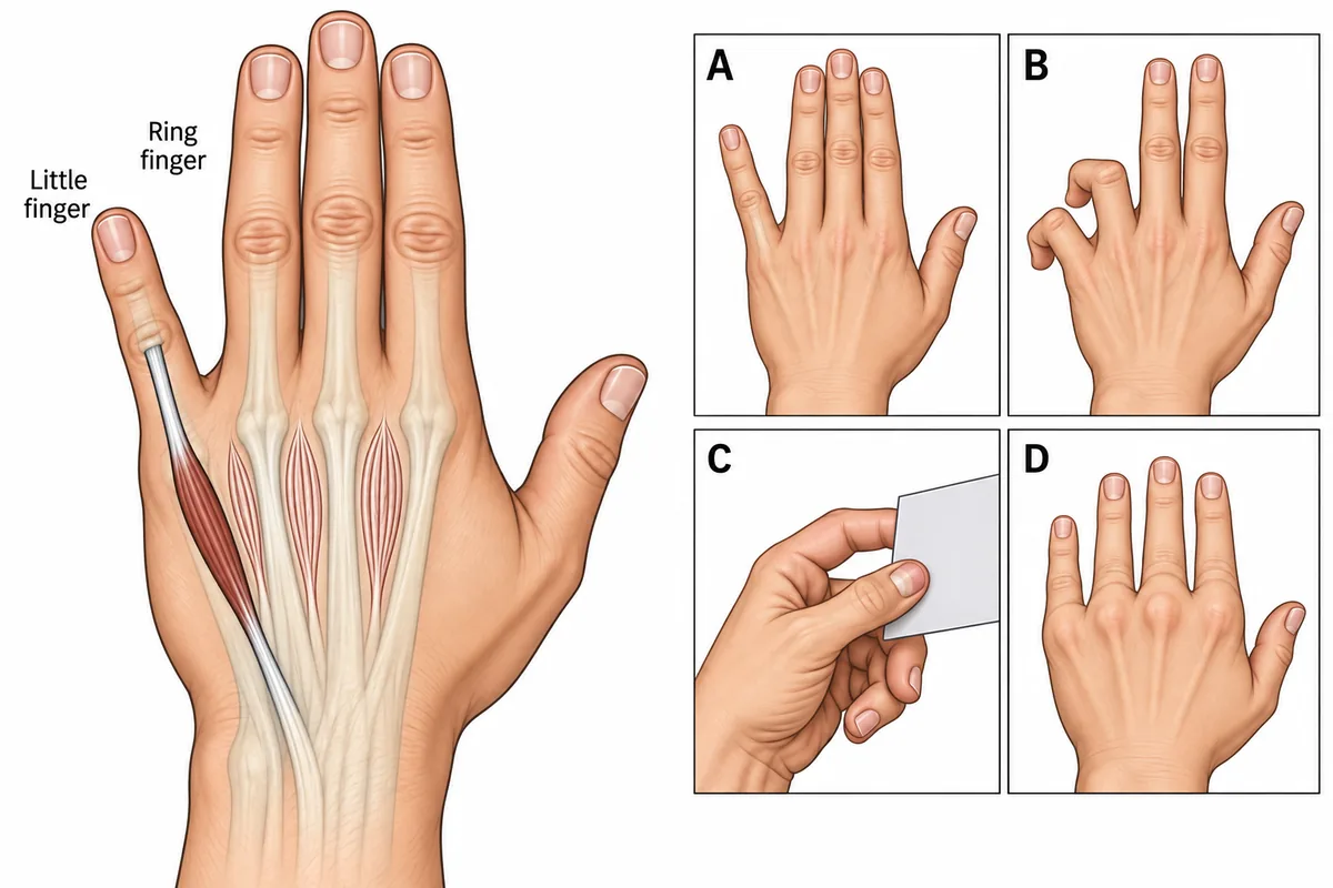
A hand is drawn in the figure lying at rest on a table, with the small muscles between the metacarpals and the long tendon to the little finger drawn in different shades and four postures lettered on separate panels. Loss of which single muscle produces the posture the resting hand shows? One hand at rest on a table with the small muscles of the palm shaded, and four postures drawn on separate panels.
Look first: count how many fingers are out of line at complete rest, then ask which muscle pulls that finger back towards the middle finger.
 View larger ↗⤢ Tap to enlarge
- AThird and fourth lumbricals
- BAdductor pollicis
- CThird palmar interosseous
- DExtensor digiti minimi
Show the answer and explanation
Answer: C. Third palmar interosseous
C. Third palmar interosseous. Only the little finger is out of line; the other three lie together. Palmar interossei pull the fingers towards the middle finger, and dorsal interossei push them apart. The third palmar interosseous is the only adductor the little finger has. Without it, the long extensor, which pulls slightly outwards, acts unopposed. The posture is present at complete rest, which separates it from a weak grip.
Why the other options lose: Third and fourth lumbricals (A): their loss gives the clawing of panel B, and that needs the lumbricals and interossei of two fingers to fail, not one small muscle. Adductor pollicis (B): its loss gives panel C, where the thumb buckles because a long flexor substitutes for the lost adductor. Extensor digiti minimi (D) is the muscle doing the pulling, so losing it would correct the drift rather than cause it.
Recallab question 56601 · Anatomy › Upper Limb · reference: Snell's Clinical Anatomy by Regions
Recallab example 2 · more damage, less deformity
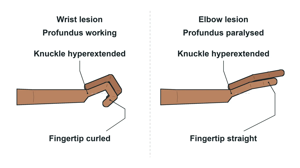
Why does a high ulnar nerve lesion at the elbow produce a LESS obvious claw hand than a low lesion at the wrist?
Look first: decide which long flexor the elbow lesion adds to the paralysis, and what that flexor does to the fingertips.
- ASparing of the lumbricals
- BParalysis of flexor digitorum profundus
- CMedian nerve compensation
- DPurely sensory loss
Show the answer and explanation
Answer: B. Paralysis of flexor digitorum profundus
 View larger ↗⤢ Tap to enlarge
B. Paralysis of flexor digitorum profundus. The higher the lesion, the less the claw. The ulnar half of flexor digitorum profundus bends the terminal joints of the ring and little fingers. A wrist lesion spares it, so those fingers flex strongly and claw sharply. An elbow lesion paralyses it too, so the terminal joints cannot curl and the claw is blunted. The deformity looks milder while the injury is worse.
Why the other options lose: Sparing of the lumbricals (A) is wrong: the ulnar lumbricals are paralysed in both lesions, which is why the knuckles hyperextend in each. Median nerve compensation (C) does not happen; the median nerve supplies the radial two lumbricals and cannot substitute. Purely sensory loss (D) is false: a high lesion causes more motor loss, not less.
Recallab question 50306 · Anatomy › Upper Limb · reference: Gray's Anatomy for Students
Where along the nerve: the wrist, the palm and the forearm
Recallab example 3 · cannot spread the fingers, can feel
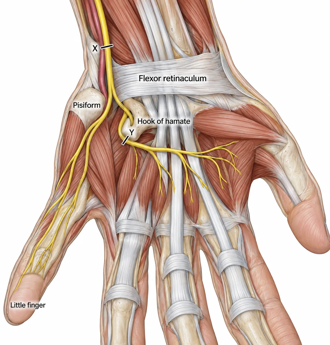
Two men cut the front of the right wrist and palm on broken glass, at the points marked X and Y in the figure. One of them can no longer spread or close his fingers, yet feels a pin on the palmar skin of the little finger normally. What did his cut divide? The ulnar side of the right wrist and palm with two cut sites marked.
Look first: find where the nerve divides, then match each cut to the branches it crosses.
 View larger ↗⤢ Tap to enlarge
- AMedian nerve at the wrist
- BSuperficial branch of the ulnar nerve
- CDeep branch of the ulnar nerve
- DUlnar nerve trunk above the pisiform
Show the answer and explanation
Answer: C. Deep branch of the ulnar nerve
C. Deep branch of the ulnar nerve. Spreading and closing the fingers is the work of the interossei, supplied by the deep branch. Cut Y crosses the deep branch at the hamate and spares the superficial branch, which carries palmar sensation from the little finger, so that skin still feels. In the palm the ulnar nerve splits by job.
Why the other options lose: The median nerve at the wrist (A) supplies no interosseous muscle and no skin of the little finger. Losing the superficial branch of the ulnar nerve (B) numbs the skin and spares the interossei, the reverse of his picture. Cut X divides the ulnar nerve trunk above the pisiform (D) before it branches, so both branches go and the palmar skin of the little finger would be numb.
Recallab question 55702 · Anatomy › Upper Limb · reference: Snell's Clinical Anatomy by Regions
Recallab example 4 · the one muscle above the aponeurosis
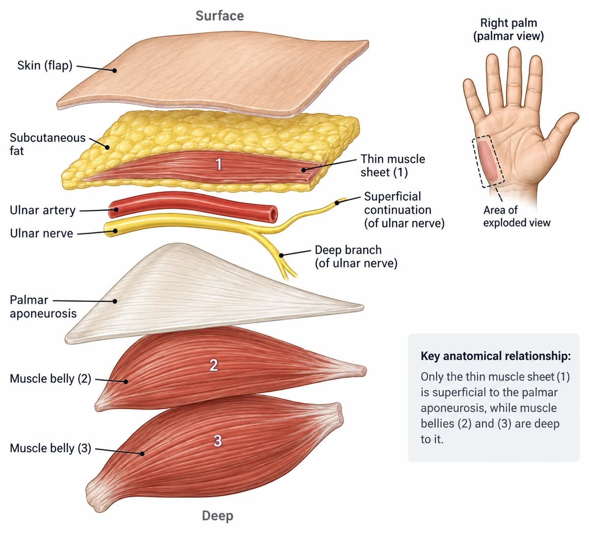
The ulnar border of the palm is drawn in the figure with the skin turned back and three muscles numbered, one of them lying in the fatty layer above the palmar aponeurosis and the other two below it. Which nerve supplies muscle 1? The ulnar border of the palm with the skin turned back and three muscles numbered.
Look first: place muscle 1 in its layer first, then follow the branch that runs in that layer.
 View larger ↗⤢ Tap to enlarge
- ADeep branch of the ulnar nerve
- BPalmar cutaneous branch of the ulnar nerve
- CRecurrent branch of the median nerve
- DSuperficial branch of the ulnar nerve
Show the answer and explanation
Answer: D. Superficial branch of the ulnar nerve
D. Superficial branch of the ulnar nerve. Muscle 1 is palmaris brevis, the only muscle of the hand in the superficial fascia, above the palmar aponeurosis. It wrinkles the skin of the ulnar border. It is the only muscle the superficial branch supplies; the rest of that branch is sensory, running on as the digital nerves of one and a half fingers.
Why the other options lose: The deep branch of the ulnar nerve (A) supplies the hypothenar bellies below the aponeurosis and the interossei, not the fatty layer above it. The palmar cutaneous branch of the ulnar nerve (B) is purely sensory to the skin over the hypothenar eminence. The recurrent branch of the median nerve (C) supplies the thenar muscles on the other side of the palm.
Recallab question 58601 · Anatomy › Upper Limb · reference: Gray's Anatomy for Students
Recallab example 5 · hand muscles spared after an elbow cut
A man's ulnar nerve was cut cleanly just above the elbow and has been repaired. Three days later his first dorsal interosseous and adductor pollicis still contract strongly, although flexor carpi ulnaris is paralysed. Four patterns of connection between the median and ulnar nerves are drawn in the figure, each with the same cut. Which drawing explains his hand? Four drawings of the median and ulnar nerves of one upper limb from the front, each with the same cut and a different connecting branch.
Look first: check the direction of each connecting arrow and whether the join lies above or below the cut.
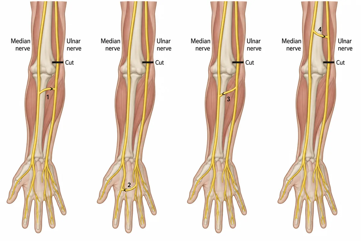 View larger ↗⤢ Tap to enlarge
- ADrawing 2
- BDrawing 4
- CDrawing 3
- DDrawing 1
Show the answer and explanation
Answer: D. Drawing 1
D. Drawing 1. Flexor carpi ulnaris is paralysed, so the ulnar nerve really is cut. The spared ulnar hand muscles must therefore get their fibres without passing the cut. In drawing 1 those fibres travel in the median nerve past the elbow and cross to the ulnar nerve in the upper forearm, below the injury. This median-to-ulnar crossing in the forearm is the Martin-Gruber anastomosis, found in roughly one limb in five.
Why the other options lose: Drawing 2 (A) is a palmar connection, which usually carries ulnar fibres towards the thenar muscles; it cannot keep the ulnar hand muscles working here. Drawing 4 (B) joins the ulnar nerve above the cut, so it is severed with it. In drawing 3 (C) the arrow runs the wrong way: fibres leaving the ulnar nerve have already passed through the cut.
Recallab question 60001 · Anatomy › Upper Limb · reference: Gray's Anatomy for Students
Timing the injury on a needle study
Recallab example 6 · three weeks after a complete crush
A man had a complete crush injury of the ulnar nerve at the elbow three weeks ago. Needle electromyography from four different muscles, each at rest and on effort, is drawn in the figure as panels 1 to 4. Which panel comes from his first dorsal interosseous? Needle electromyography from four muscles, each at rest and on effort, numbered 1 to 4.
Look first: read the at-rest strip first: a denervated muscle is not silent at rest.
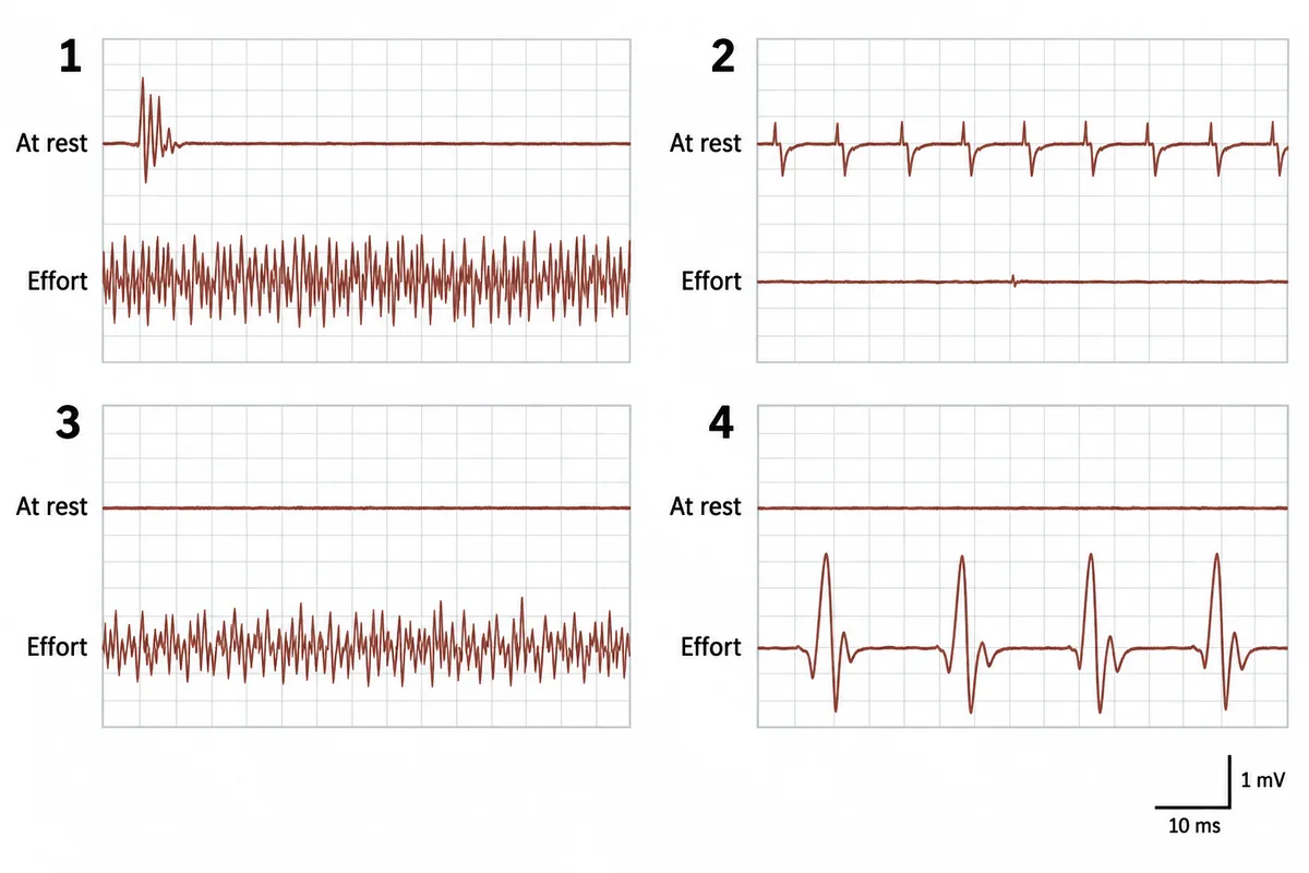 View larger ↗⤢ Tap to enlarge
- APanel 2
- BPanel 1
- CPanel 3
- DPanel 4
Show the answer and explanation
Answer: A. Panel 2
A. Panel 2. The first dorsal interosseous is supplied by the ulnar nerve, and a complete crush cuts every axon. Denervated fibres become hypersensitive and fire on their own: fibrillation potentials are tiny regular spikes, and positive sharp waves point downwards with a slow return. They take two to three weeks to appear, the time for Wallerian degeneration to reach the muscle. With no axons, almost no motor units appear on effort.
Why the other options lose: Panel 1 (B), with silence at rest and a full interference pattern on effort, is normal muscle. Panel 3 (C), with small polyphasic units recruited early, is the pattern of a myopathy, and the nerve crush has spared the muscle fibres. Panel 4 (D), with large units and reduced recruitment, is chronic reinnervation, which takes months, not three weeks.
Recallab question 55509 · Physiology › Nerve & Muscle · reference: Guyton and Hall Textbook of Medical Physiology
The one-minute routine
| If the figure gives | Ask first | It settles |
|---|---|---|
| One finger out of line at rest | Which muscle pulls it back towards the middle finger? | The little finger drifts out when its palmar interosseous is lost |
| A claw that looks mild after a high lesion | Is flexor digitorum profundus working? | Profundus paralysed blunts the claw: more damage, less deformity |
| Weak interossei with normal palmar skin | Which branch does the cut cross? | The deep branch moves; the superficial branch mostly feels |
| A thin muscle above the palmar aponeurosis | Which layer is it in? | Palmaris brevis is the superficial branch's only muscle |
| Hand muscles spared after an elbow cut | Where do the fibres cross, and which way? | Median to ulnar, in the forearm, below the lesion |
| Spontaneous spikes at rest weeks after an injury | How long since the injury? | Fibrillations and sharp waves appear at two to three weeks |
Practise anatomy questions and see the same reasoning across the nervous system in neurology localisation. For other cut surfaces, see anatomy cross-sections.
Why candidates use Recallab
- Built towards 22,000 — a batch at a time
- Illustrations drawn for the question
- Every option explained, not just the right one
- AI study tools that are live today
- Start free, today
Or see the plans, or try a timed mock section.
Register to practise NEET PG anatomy questionsFree account, no card needed. Practise with every option explained and a reference on every question.Drawn from questions #56601, #50306, #55702, #58601, #60001, #55509 in the Recallab bank, each with its own verified answer and current-edition reference. Last reviewed 25 October 2026.