
Recallab › Blog › Biochemistry for NEET PG: six pathways and the one blocked step in each
Biochemistry for NEET PG: six pathways and the one blocked step in each
A pathway question is not a memory test of the whole map. Find the one blocked step, and what piles up behind it tells you the illness.
Register to practise NEET PG biochemistry questions →Free account, no card needed.
These questions draw a pathway, number its steps and describe a patient. The pictures below are illustrations drawn for the questions. Each is a real Recallab MentorQ, with the reasoning that decides it. Try each before you open the answer.
Before you pick a step: three questions
- What has piled up? The compound named in the stem sits just before the block.
- What is missing downstream? A product that is absent, such as ketones, points back to the step that should have made it.
- Which organ is hurt, and by what? The damage often comes from a side product of the block, not from the block itself.
A sugar and two amino acid pathways
Recallab example 1 · a milk-fed baby
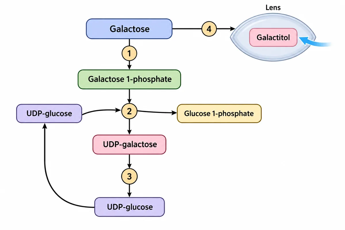
A two-week-old breastfed baby has vomiting, jaundice, an enlarged liver, a metabolic acidosis and early cataracts. The pathway of galactose is drawn in the figure with four enzyme steps numbered, and step 2 is blocked. Which accumulating compound injures the liver and kidney?
Look first: ask which compound sits just above each numbered step, and where each one collects.
 View larger ↗⤢ Tap to enlarge
- AUDP-galactose
- BGalactitol
- CGalactose 1-phosphate
- DFree galactose
Show the answer and explanation
Answer: C. Galactose 1-phosphate
C. Galactose 1-phosphate. Step 2 is the uridyltransferase, which uses galactose 1-phosphate to form UDP-galactose. When it is blocked, galactose 1-phosphate builds up inside cells and injures the liver and kidney tubules. The ill milk-fed baby has classic galactosaemia. Galactose also backs up and is reduced to galactitol in the lens, where it draws in water and causes cataracts.
Why the other options lose: UDP-galactose (A) is the product of the blocked step, not the compound building up behind it. Galactitol (B) explains the cataracts, not the liver and kidney injury. Free galactose (D) also rises, but galactose 1-phosphate is the damaging compound asked for here. Keep the two sites apart: galactose 1-phosphate in liver and kidney, galactitol in the lens.
Recallab question 55811 · Biochemistry › Carbohydrate Metabolism · reference: Harper's Illustrated Biochemistry
Recallab example 2 · a newborn with high ammonia
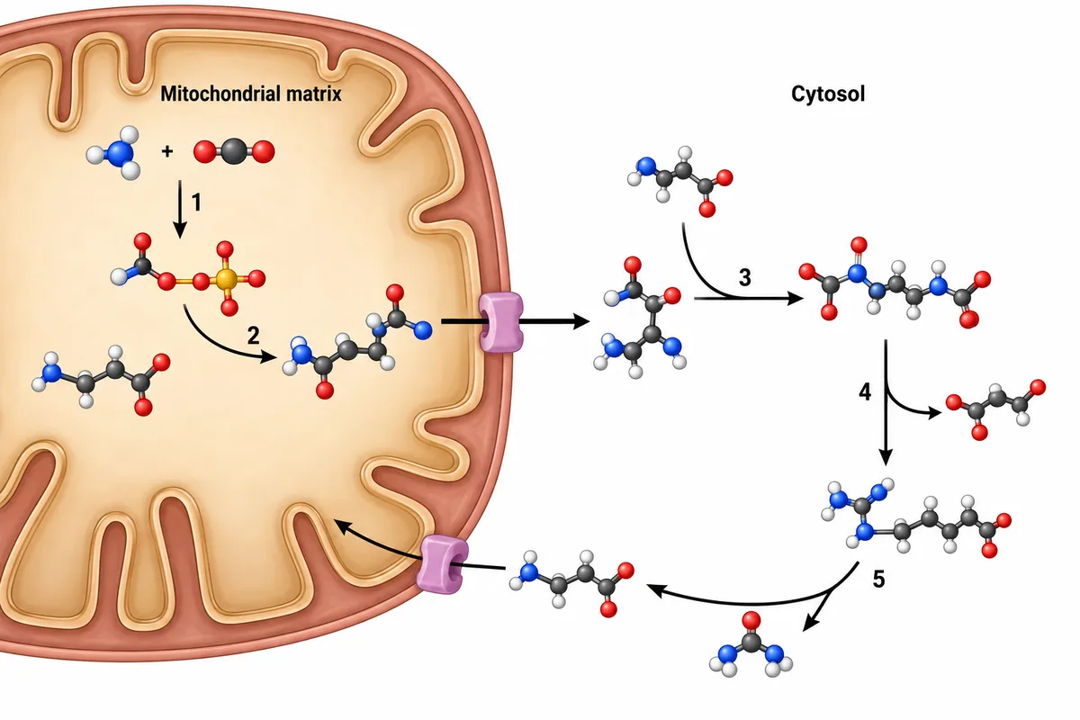
The urea cycle is drawn in the figure across the mitochondrial membrane with its five enzyme steps numbered. A neonate has hyperammonaemia, a markedly raised plasma citrulline and a normal urinary orotic acid. Which numbered step is blocked?
Look first: find where citrulline is made and where it is used, then ask what orotic acid tells you.
 View larger ↗⤢ Tap to enlarge
- AStep 1, carbamoyl phosphate synthetase I
- BStep 3, argininosuccinate synthetase
- CStep 4, argininosuccinate lyase
- DStep 2, ornithine transcarbamylase
Show the answer and explanation
Answer: B. Step 3, argininosuccinate synthetase
B. Step 3, argininosuccinate synthetase. Citrulline is made at step 2 inside the mitochondrion and carried into the cytosol. Step 3 joins it to aspartate to make argininosuccinate. A block here lets citrulline build up sharply: citrullinaemia type I. The normal urinary orotic acid in this stem helps distinguish the pattern from an ornithine transcarbamylase block.
Why the other options lose: step 1, carbamoyl phosphate synthetase I (A), lowers citrulline because less carbamoyl phosphate is made. Step 4, argininosuccinate lyase (C), raises argininosuccinic acid. Step 2, ornithine transcarbamylase (D), gives low citrulline and raised orotic acid as unused carbamoyl phosphate enters pyrimidine synthesis. Markedly raised citrulline points to step 3.
Recallab question 55311 · Biochemistry › Amino Acid Metabolism · reference: Harper's Illustrated Biochemistry
Recallab example 3 · a marker in the urine
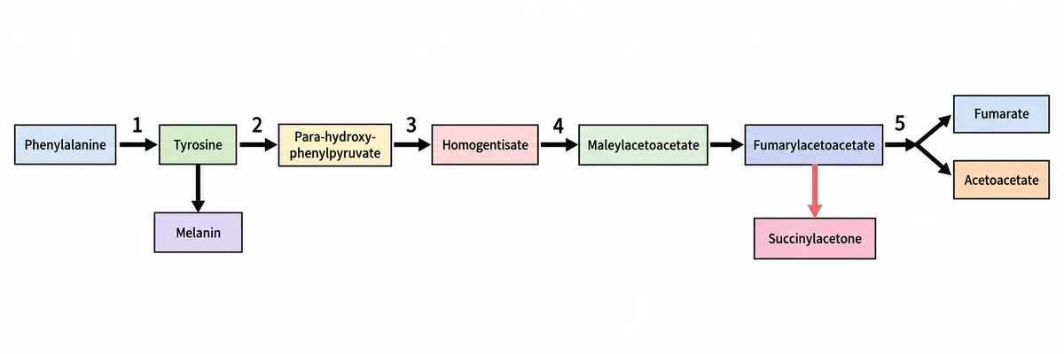
The breakdown of phenylalanine and tyrosine is drawn in the figure with five enzyme steps numbered. An infant has liver failure and renal Fanconi syndrome, and succinylacetone is found in the urine. Which numbered step is deficient?
Look first: find the compound named in the stem on the figure, and see which box it hangs from.
 View larger ↗⤢ Tap to enlarge
- AStep 2, tyrosine aminotransferase
- BStep 5, fumarylacetoacetate hydrolase
- CStep 4, homogentisate oxidase
- DStep 1, phenylalanine hydroxylase
Show the answer and explanation
Answer: B. Step 5, fumarylacetoacetate hydrolase
B. Step 5, fumarylacetoacetate hydrolase. This is tyrosinaemia type I. A block at the last step backs up fumarylacetoacetate, with succinylacetone formed along the side branch. These compounds damage liver cells and the proximal kidney tubule, explaining liver failure with renal Fanconi syndrome. Succinylacetone in urine or blood is the marker of this defect.
Why the other options lose: step 2, tyrosine aminotransferase (A), causes tyrosinaemia type II, which affects the eyes and skin. Step 4, homogentisate oxidase (C), causes alkaptonuria, with dark urine and later ochronosis. Step 1, phenylalanine hydroxylase (D), causes phenylketonuria, with a risk of intellectual disability if untreated. None explains the succinylacetone marker. Liver and kidney injury with that marker points to step 5.
Recallab question 55411 · Biochemistry › Amino Acid Metabolism · reference: Harper's Illustrated Biochemistry
Two fat pathways and haem
Recallab example 4 · low glucose, no ketones
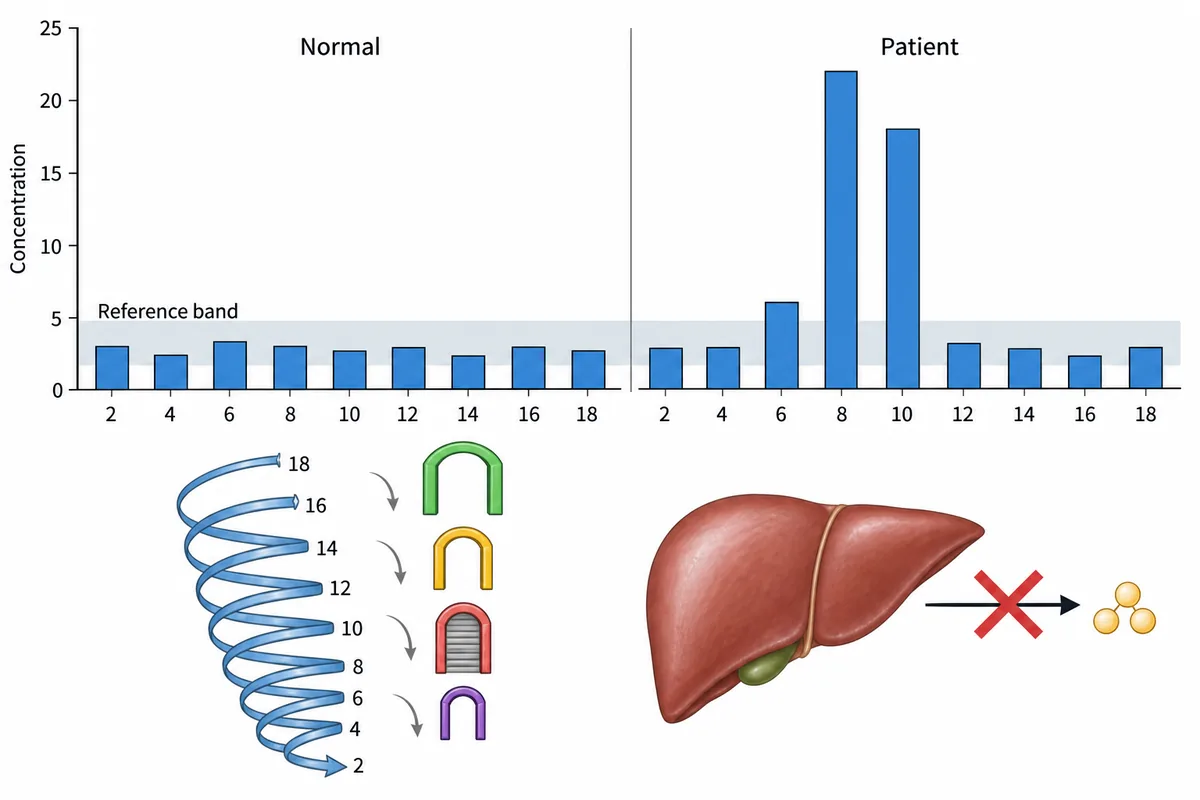
A well infant was found unrousable one morning after a long night without a feed, with a blood glucose of 29 mg/dL (1.6 millimoles per litre) and no ketones in her urine. Her dried blood spot profile is drawn in the figure against the reference band. What does the profile show?
Look first: read along the patient's bars and note the chain lengths that stand above the band.
 View larger ↗⤢ Tap to enlarge
- ALong chain fatty acid oxidation block
- BKetotic hypoglycaemia
- CMedium chain fatty acid oxidation block
- DCarnitine uptake defect
Show the answer and explanation
Answer: C. Medium chain fatty acid oxidation block
C. Medium chain fatty acid oxidation block. The tall bars at eight and ten carbons, with a smaller rise at six, place the block at medium chain lengths. Beta oxidation normally shortens fatty acids and releases acetyl groups for ketone formation. When medium chain oxidation stalls, the liver has less substrate for ketone synthesis. A fasting infant can therefore become severely hypoglycaemic without making enough ketones.
Why the other options lose: a long chain fatty acid oxidation block (A) would raise long chain species, whereas those bars lie within the reference band. Ketotic hypoglycaemia (B) includes ketones, which are absent here. A carnitine uptake defect (D) gives low free carnitine and broadly low acylcarnitines, rather than this rise in medium chain species. The missing ketones point to a fat oxidation problem; the raised chain lengths locate it.
Recallab question 57311 · Biochemistry › Lipid Metabolism · reference: Harper's Illustrated Biochemistry
Recallab example 5 · a door into the peroxisome
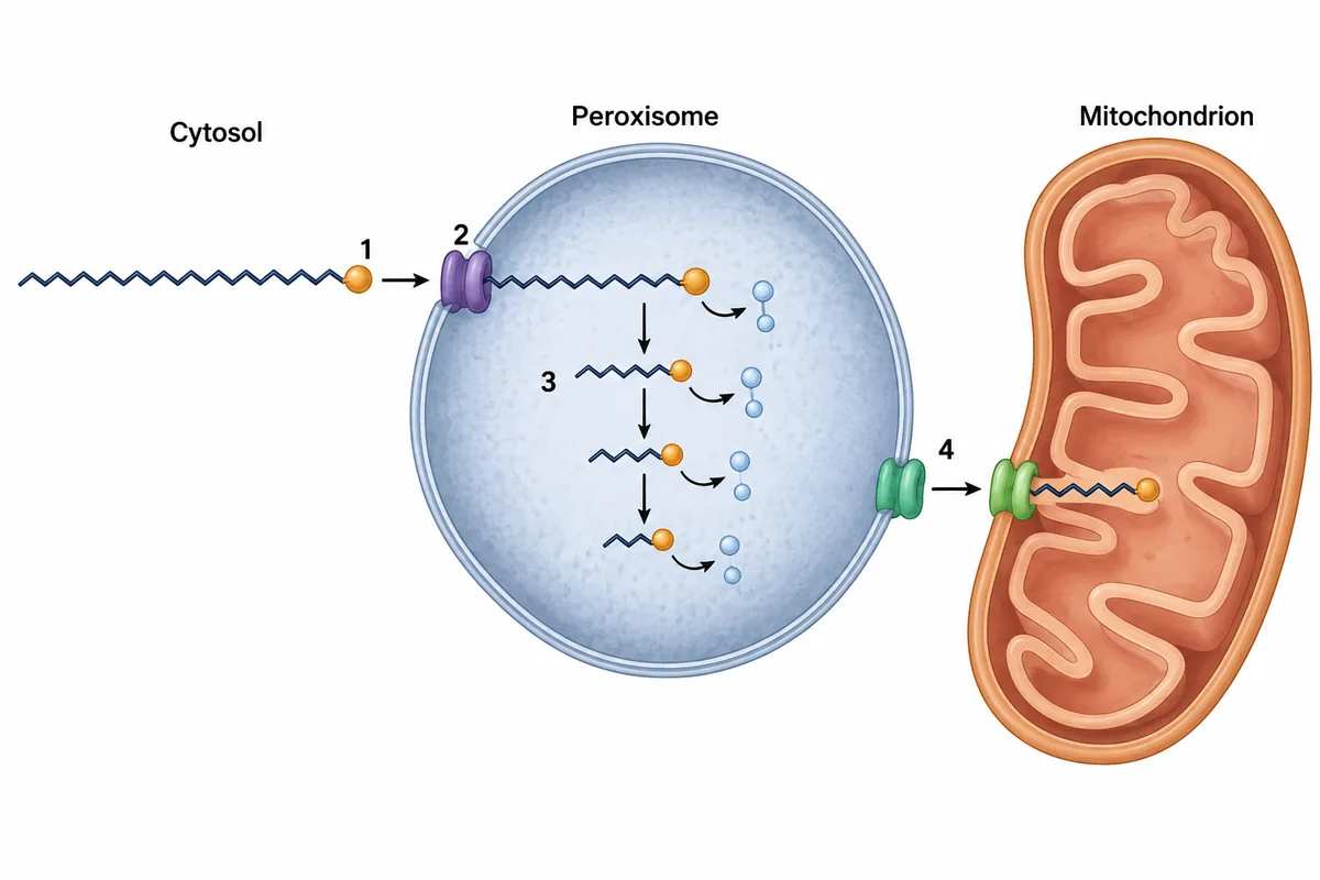
A boy of eight has falling school performance and darkening skin; his mother's brother died of a similar illness. Plasma very long chain fatty acids are high, and his peroxisomes are present and look normal. The figure traces such a fatty acid through a peroxisome, with four steps numbered. Which numbered step has failed?
Look first: follow the chain from left to right and name what each numbered step does to it.
 View larger ↗⤢ Tap to enlarge
- AStep 1
- BStep 3
- CStep 2
- DStep 4
Show the answer and explanation
Answer: C. Step 2
C. Step 2. This is the transporter at the peroxisomal membrane that allows very long chain fatty acids to enter for shortening. Its gene lies on the X chromosome. Failure of transport lets these fatty acids build up, damaging the adrenal cortex and the white matter of the brain. Darkening skin and falling school performance, with an affected maternal uncle, fit X-linked adrenoleukodystrophy. The peroxisomes themselves are present.
Why the other options lose: step 1 (A) activates the fatty acid in the cytosol; it is not the membrane transport step linked to this pattern. Step 3 (B) shortens the chain inside the peroxisome. Defects there can also raise very long chain fatty acids, but do not explain the characteristic X-linked pattern as well. Step 4 (D) carries the shortened chain into the mitochondrion for further oxidation. The adrenal and white matter clues locate the failed door at step 2.
Recallab question 56013 · Biochemistry › Lipid Metabolism · reference: Harper's Illustrated Biochemistry
Recallab example 6 · two blocks in one pathway
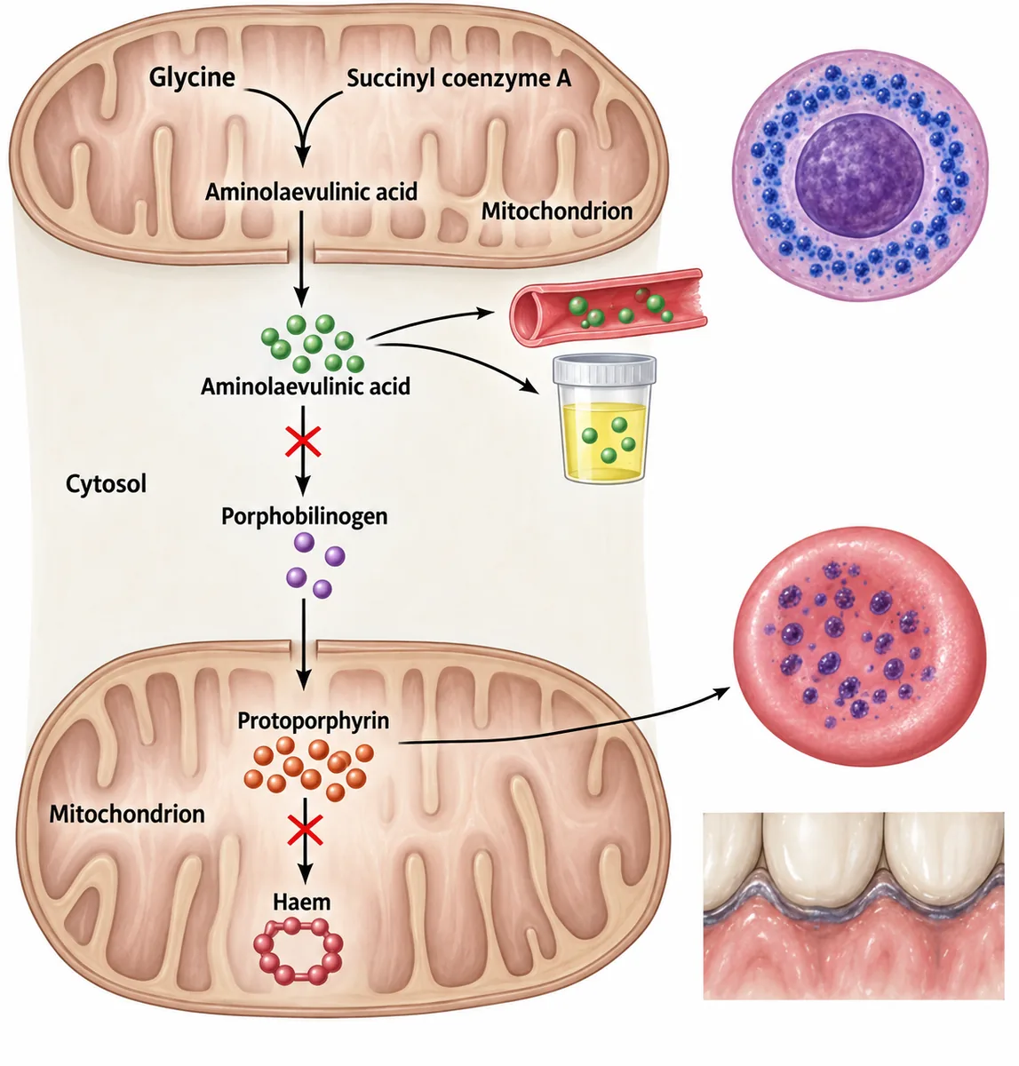
The haem synthesis pathway is drawn in the figure with two steps crossed out and the metabolites that accumulate shown. What will the blood film show?
Look first: count the crossed-out steps, then note which compound collects above each cross.
 View larger ↗⤢ Tap to enlarge
- AHypersegmented neutrophils
- BMicrocytes with pencil cells
- CTarget cells and acanthocytes
- DCoarse basophilic stippling
Show the answer and explanation
Answer: D. Coarse basophilic stippling
D. Coarse basophilic stippling. Lead blocks two enzymes of haem synthesis: the one that uses aminolaevulinic acid and the one that inserts iron into protoporphyrin at the end. Aminolaevulinic acid and protoporphyrin therefore accumulate. Lead also inhibits the breakdown of ribosomal RNA in young red cells. The remaining RNA clumps into coarse blue granules, giving the blood film finding shown.
Why the other options lose: hypersegmented neutrophils (A) point to megaloblastic anaemia. Microcytes with pencil cells (B) suggest iron deficiency; lead can cause small red cells, but coarse stippling is the finding that fits this pathway picture. Target cells and acanthocytes (C) can occur in liver disease and do not explain the two blocked haem enzymes. Two pathway blocks with coarse blue granules point to lead and option D.
Recallab question 55113 · Biochemistry › Vitamins & Minerals · reference: Harper's Illustrated Biochemistry
The one-minute routine
| If the stem gives | Ask first | It usually settles |
|---|---|---|
| A milk-fed baby with jaundice and cataracts | Is the baby ill, or is it cataract alone | Transferase block or kinase block |
| A newborn with high ammonia | Is citrulline raised or low, and is orotic acid raised | Which urea cycle step is blocked |
| Liver failure with a renal tubular defect | Is succinylacetone present | Tyrosinaemia type I |
| Low glucose after a fast | Are there ketones | A fat oxidation defect if there are none |
| Raised very long chain fatty acids | Are the peroxisomes present, and who else is affected | A transporter defect or absent peroxisomes |
| Two blocks in haem synthesis | Which compounds collect above each block | Lead |
For other kinds of figure, see ten NEET PG image questions and acid-base and electrolytes. For curves and plots, see physiology graphs.
Why candidates use Recallab
- Built towards 22,000 — a batch at a time
- Illustrations drawn for the question
- Every option explained, not just the right one
- AI study tools that are live today
- Start free, today
Or see the plans, or try a timed mock section.
Register to practise NEET PG biochemistry questionsFree account, no card needed. Practise with every option explained and a reference on every question.Drawn from questions #55811, #55311, #55411, #57311, #56013, #55113 in the Recallab bank, each with its own verified answer and current-edition reference. Last reviewed 12 October 2026.