
Recallab › Blog › Radiology for NEET PG: six plain films and the rule that reads each one
Radiology for NEET PG: six plain films and the rule that reads each one
A plain film is read with rules, not hunches. Learn one rule for each film and the picture does most of the work.
Register to practise NEET PG radiology questions →Free account, no card needed.
These questions show a drawn radiograph and ask what it shows. The pictures below are illustrations drawn for the questions, not patient films. Each is a real Recallab MentorQ, with the rule that decides it. Try each before you open the answer.
Before you name a film: three questions
- What am I looking at? Name the view and the region, so you know what normal looks like.
- What is the one thing out of place? A shadow, a line, a gas pocket, a bone that is too white.
- Which rule applies? Most plain-film signs come with a rule: which border is lost, which line should cross which bone.
The chest and the abdomen
Recallab example 1 · a collapsed lobe
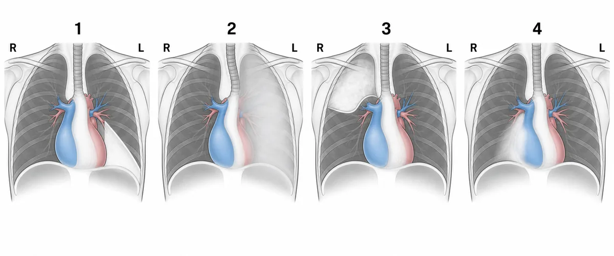
Four frontal chest radiographs, each from a patient with one lobe collapsed, are drawn in the figure. Which drawing shows collapse of the left lower lobe?
Look first: ask which border or outline is lost in each drawing, and which lobe touches it.
 View larger ↗⤢ Tap to enlarge
- ADrawing 1
- BDrawing 3
- CDrawing 4
- DDrawing 2
Show the answer and explanation
Answer: A. Drawing 1
Drawing 1. The collapsed left lower lobe folds down and back against the spine and the diaphragm. Seen from the front it forms a dense triangle behind the heart, the sail sign, whose outer edge is the displaced oblique fissure. Because the lobe touches the diaphragm, the inner left diaphragm outline is lost, while the left heart border stays sharp. The left hilum is pulled down by the loss of volume.
Why the closest rival loses: drawing 2 (D) is a veil over the whole left lung with the left heart border lost and the trachea pulled to the left: collapse of the left upper lobe. Drawing 4 (C) loses the right heart border, which is right middle lobe disease. Drawing 3 (B) is an opacity in the right upper zone, right upper lobe collapse. Lower lobes hide the diaphragm; the lingula and the middle lobe hide the heart borders.
Recallab question 55745 · Radiology › Chest Imaging · reference: Harrison's Principles of Internal Medicine
Recallab example 2 · gas under the diaphragm
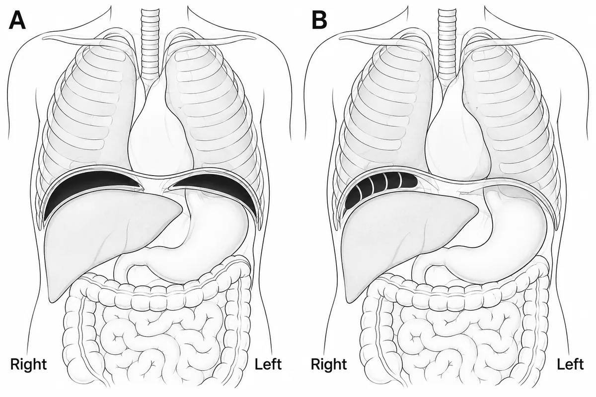
Two erect chest films are drawn in the figure, each showing gas beneath the right side of the diaphragm. One of them is a harmless look-alike of perforation. Which feature identifies it?
Look first: look inside each gas pocket for lines, and at which sides the gas lies.
 View larger ↗⤢ Tap to enlarge
- AAn air-fluid level
- BGas under both domes
- CHaustral folds within the gas
- DGas confined to the right side
Show the answer and explanation
Answer: C. Haustral folds within the gas
Haustral folds within the gas. Free gas under the diaphragm is a smooth crescent with no internal lines. In film B, lines cross the gas pocket, and those are haustral folds of bowel wall. They prove the gas lies inside a loop of colon that has slipped between the liver and the right diaphragm: a benign anatomical variant, the Chilaiditi sign. Recognising the folds avoids an unnecessary laparotomy for presumed perforation.
Why the closest rival loses: gas under both domes (B) is film A, the true free-gas pattern. Gas confined to the right side (D) does not decide it, because free gas from a perforated ulcer can be right-sided too. An air-fluid level (A) suggests an abscess, and neither film shows one. Lines crossing the gas mean bowel wall, not a hole in the gut.
Recallab question 55681 · Radiology › Abdominal & GI Imaging · reference: Bailey & Love's Short Practice of Surgery
Recallab example 3 · a loop that stands guard
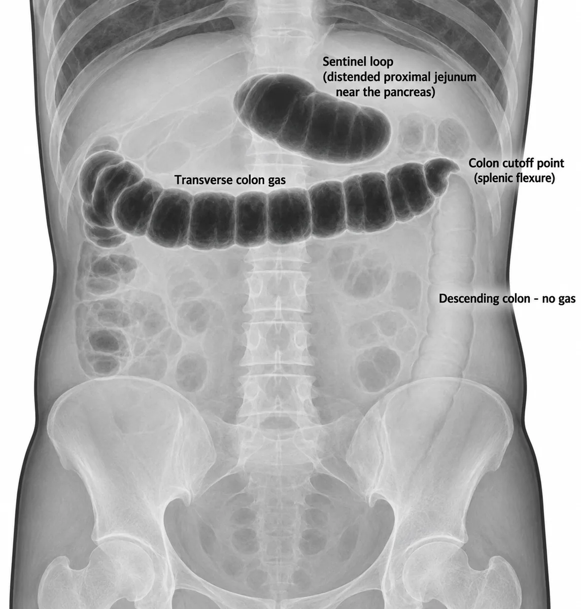
A plain abdominal film is drawn in the figure with gas distending a single loop of proximal jejunum near the pancreas, and gas in the transverse colon ending abruptly at the splenic flexure with none seen beyond it. What condition does this combination point to?
Look first: count the dilated loops, then look for a point where the bowel narrows.
 View larger ↗⤢ Tap to enlarge
- ALarge bowel obstruction
- BA normal gas pattern
- CSmall bowel obstruction
- DAcute pancreatitis
Show the answer and explanation
Answer: D. Acute pancreatitis
Acute pancreatitis. A single distended loop near the pancreas is a sentinel loop: a localised reaction to nearby inflammation. Spasm of the colon from the same inflammation halts gas at the splenic flexure, the colon cutoff sign. No transition point or narrowing is drawn, which argues against a true mechanical block. The finding supports pancreatitis rather than proves it.
Why the closest rival loses: a large bowel obstruction (A) needs a transition point or narrowing, and none is drawn. A small bowel obstruction (C) usually shows several dilated loops, not one. A normal gas pattern (B) does not contain a distended loop with an abrupt cutoff. Sentinel loop plus colon cutoff, without a transition point, means inflammation, not obstruction.
Recallab question 56045 · Radiology › Abdominal & GI Imaging · reference: Harrison's Principles of Internal Medicine
Bones and joints
Recallab example 4 · a line on the skull
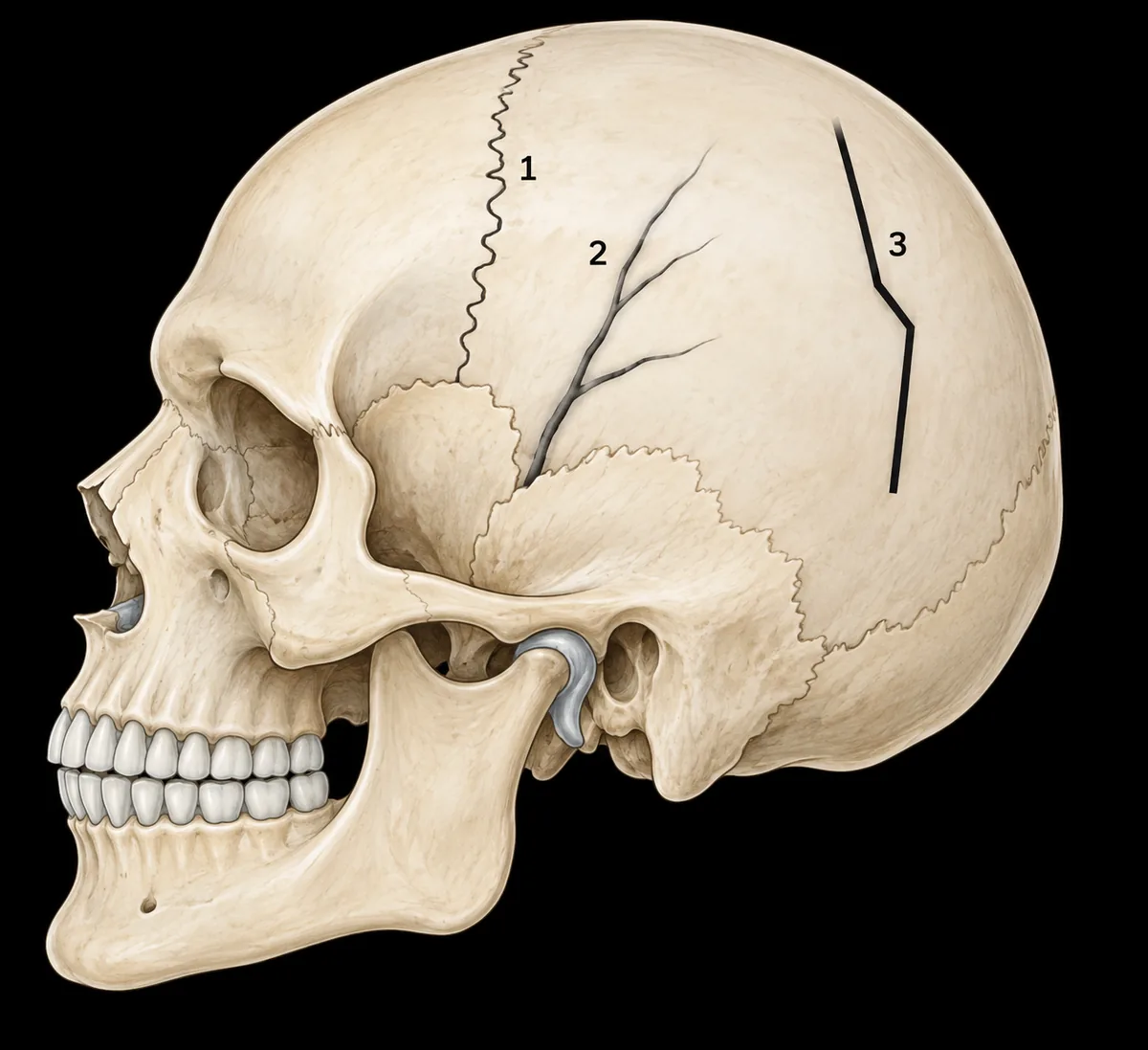
A 6-year-old fell from a first-floor balcony. A lateral skull radiograph is drawn in the figure with three lucent lines numbered. Which line is a fracture?
Look first: describe each line: how dark, how straight, whether it branches and what its edges look like.
 View larger ↗⤢ Tap to enlarge
- ALine 3
- BLine 2
- CLine 1
- DLines 2 and 3
Show the answer and explanation
Answer: A. Line 3
Line 3. A linear skull fracture passes through both tables, so it looks blacker than a groove. It is sharply lucent, straight or angular, unbranched and has no white margin, because there has been no time for a sclerotic rim to form. It also follows no anatomical course. Line 3 shows every one of those features.
Why the closest rival loses: line 2 (B) branches and tapers with smooth, white-edged margins along the course of a meningeal vessel: a vascular groove. Line 1 (C) zigzags with white margins where the coronal suture lies: a normal suture. Lines 2 and 3 together (D) is wrong, because a groove is a normal impression, not a second fracture. Fracture: black, straight, bare-edged. Suture: zigzag. Groove: branching.
Recallab question 58045 · Radiology › Skeletal System · reference: Bailey & Love's Short Practice of Surgery
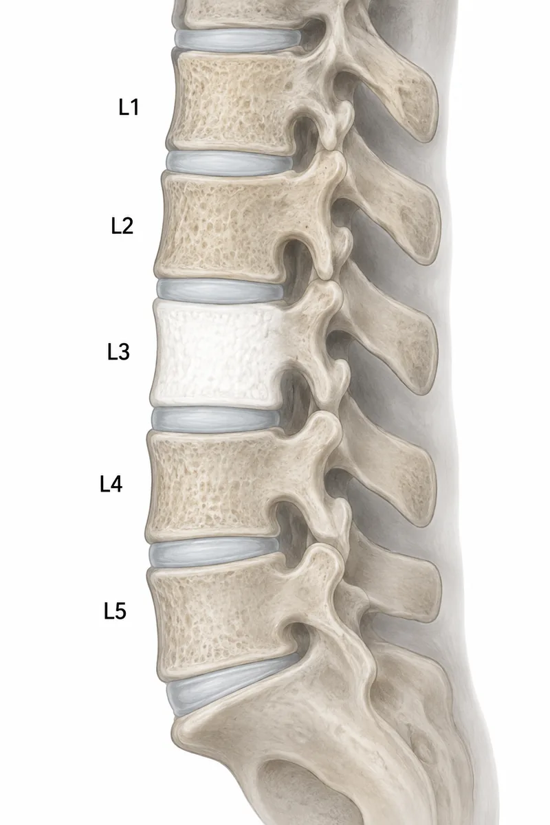
Recallab example 5 · a white vertebra
A man of 70 has had low back pain for two months. His lateral lumbar spine radiograph is drawn in the figure. What is the most likely cause of the abnormal vertebra?
Look first: compare the shade of each vertebra, then compare its size and outline with its neighbours.
 View larger ↗⤢ Tap to enlarge
- AOsteoporotic compression fracture
- BPaget disease
- COsteoblastic metastasis
- DVertebral haemangioma
Show the answer and explanation
Answer: C. Osteoblastic metastasis
Osteoblastic metastasis. L3 is uniformly white, yet its size and outline match its neighbours. A uniformly dense vertebra of normal size is an ivory vertebra, which in an adult points to sclerotic metastasis or lymphoma. In an older man, prostate cancer is the commonest source of sclerotic deposits. The prostate-specific antigen and a bone scan are the next steps.
Why the closest rival loses: Paget disease (B) also enlarges the body and thickens its rim, and neither is drawn. An osteoporotic compression fracture (A) would show lost height and wedging, but L3 has kept its full height. A haemangioma (D) gives coarse vertical stripes, not uniform density. Ivory and normal size: metastasis or lymphoma. Ivory and enlarged: Paget.
Recallab question 58145 · Radiology › Skeletal System · reference: Harrison's Principles of Internal Medicine
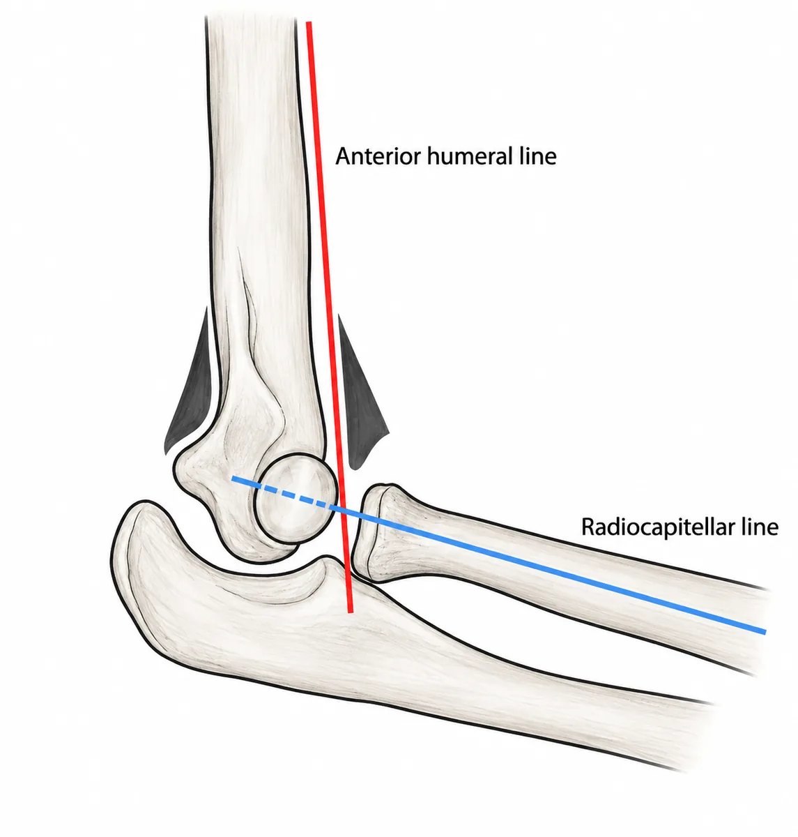
Recallab example 6 · a child's elbow
A boy of six fell on an outstretched hand. His lateral elbow radiograph is drawn with two construction lines in place. What do the lines show?
Look first: follow each line and ask which bone it should cross, and where.
 View larger ↗⤢ Tap to enlarge
- AA pulled elbow
- BA dislocated radial head
- CA normal elbow
- DA supracondylar fracture
Show the answer and explanation
Answer: D. A supracondylar fracture
A supracondylar fracture. The anterior humeral line should pass through the middle third of the capitellum. Here it passes in front of it, which means the capitellum has moved backward relative to the shaft: the distal fragment has tilted back, the usual extension-type injury. The radiocapitellar line is normal, so the radial head is not dislocated. A visible posterior fat pad is always abnormal and means blood in the joint.
Why the closest rival loses: a dislocated radial head (B) is what the radiocapitellar line detects, and here it passes correctly through the capitellum. A normal elbow (C) is wrong, because one normal line does not make the elbow normal when the anterior humeral line is displaced. A pulled elbow (A) is a subluxation of the radial head in a toddler, with normal radiographs.
Recallab question 54782 · Radiology › Musculoskeletal & Obstetric Imaging · reference: Essential Orthopaedics
The one-minute routine
| If you see | Ask first | It usually settles |
|---|---|---|
| A whitened area on a chest film | Which outline or border is lost | Which lobe has collapsed |
| Gas under the right diaphragm | Are there lines crossing the gas | Free gas or interposed colon |
| One loop of bowel distended | Is there a point where the bowel narrows | Inflammation or obstruction |
| A lucent line on the skull | Is it black, straight and bare-edged | Fracture, suture or groove |
| One vertebra a different shade | Is it still the same size as its neighbours | Metastasis or lymphoma, or Paget |
| A child's elbow with lines | Does the humeral line cross the capitellum | The fracture, and which way it tilts |
For other kinds of figure, see ten NEET PG image questions and six eye and ENT spotters. For the decision that follows the picture, see next-best-step management questions.
Why candidates use Recallab
- Built towards 22,000 — a batch at a time
- Illustrations drawn for the question
- Every option explained, not just the right one
- AI study tools that are live today
- Start free, today
Or see the plans, or try a timed mock section.
Register to practise NEET PG radiology questionsFree account, no card needed. Practise with every option explained and a reference on every question.Drawn from questions #55745, #55681, #56045, #58045, #58145, #54782 in the Recallab bank, each with its own verified answer and current-edition reference. Last reviewed 12 October 2026.